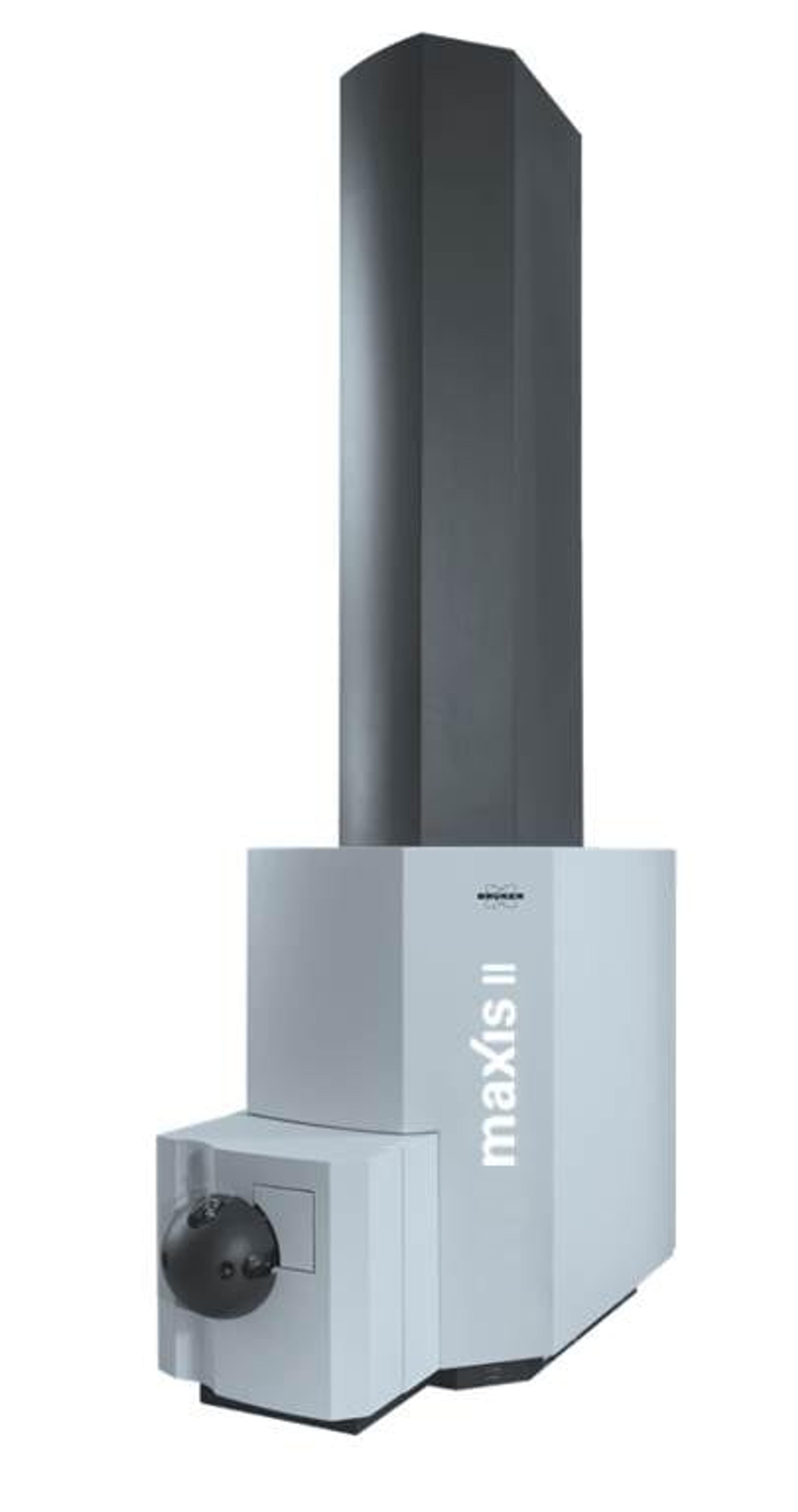
Analysis of Biosimilars and Biobetters using the maXis UHR-qTOF: A Complementary Approach
Written by Jason S. Wood, Ph.D. Bruker Daltonics
29 Mar 2015

Jason S. Wood, Ph.D. Bruker Daltonics
Biotherapeutics, specifically monoclonal antibodies, are an ever-increasing area for the pharmaceutical and biotechnology companies. Biosimilars is a term coined to describe the follow-on biologics, “generic” versions, developed after a biotherapeutic has lost patent protection. Biobetters is the term used to describe a follow-on biological with improved safety, efficacy or cost when compared to the original biotherapeutic. The main task for biosimilar developers is in proving that the primary sequence of a biosimilar is identical to its original reference material, a requirement for the regulatory process involved in getting a biosimilar approved by government agencies, even after being produced in a potentially modified biological system or from different clones.
Analysis of Biosimilars/Biobetters
Biotherapeutics already require advanced, high resolution, mass spectrometry (MS) methods for complete characterization during their initial development. Methods that include ultrahigh resolution quadrupole time-of-flight (UHR-qTOF), Fourier Transform MS (FTMS) combined with matrix assisted laser desorption (MALDI) or ESI ionization are established techniques within the biotherapeutics space due to the large size (approx. 150 kDa) and complexity (secondary, tertiary and quaternary structures) of antibodies. Simply comparing the intact mass of an antibody reference material to a biosimilar product requires advanced mass spectrometer systems and advanced techniques.
Recently, an error in the published sequence of cetuximab (a candidate for biosimilar development), seen in this article, was discovered by the use of Bruker’s maXis UHR-qTOF for analysis at the subunit level (middle-up). With this technique, advanced enzymes such as Genovis’ FabRICATOR™ (IdeS) enzyme, were used to cleave the cetuximab antibody in the conserved hinge region, allowing the antibody to be analyzed in ~25 kDa fragments, without the artifacts and time-consuming analysis required by peptide mapping [1]. To verify the identity of, and localize the amino acid substitution found in the cetuximab antibody, this work also utilized the complementary abilities of MALDI-TOF/TOF-MS on Bruker’s UltrafleXtreme. MALDI Top-down sequencing (TDS) is an excellent de novo sequencing technique that rapidly analyzes intact proteins or large fragments thereof, thus allowing targeted sequencing of medium-sized proteins, such as those generated by the FabRICATOR enzyme [2]. With this technique, a +58 Da modification in the light chain of cetuximab was found, and through the use of appropriate sequence tags an A213E amino acid substitution was determined (and confirmed by peptide mapping) to be the cause of the mass shift in the antibody. In all, two modifications were discovered in cetuximab’s sequence, when compared to the IMGT database, a cysteine (214) is missing and an alanine (213) to glutamic acid substitution was found.
Glycosylation Analysis Using MALDI
Previous work to identify the glycans of cetuximab utilized released glycans (endoglycosidase treatment) to identify and quantify the total glycans of the antibody [3]. The disadvantage of released glycan assessment is that localization of the glycan within the primary structure of the antibody is no longer possible and only an averaged glycan profile can be determined. MALDI-TOF/TOF has the advantage that the glycopeptides, obtained during a typical bottom-up analysis, can be separated and analyzed by MALDI-TOF/TOF-MS/MS with Bruker’s ProteinScape software determining the m/z value for both the peptide and glycan. Fragments are calculated and assigned to the respective MS/MS spectra. As such, a list of glycan structures is determined and ranked, with the software performing the additional task of annotating the glycans within the spectrum viewer. Importantly, the glycans can be assigned to the portion of the antibody they are attached (Fd vs. Fc), an important distinction from the use of endoglycosidases that release the glycan from the peptide backbone.
Conclusions
For a biosimilar to be developed and submitted to regulatory agencies, the primary sequence of the biosimilar must be determined and matched to the innovators original reference material. The complementary use of Bruker’s maXis UHR-qTOF system and the UltrafleXtreme MALDI-TOF/TOF MS allows for middle-up analysis of monoclonal antibodies in a fast, efficient way without the potential artifacts introduced by peptide mapping. Additionally, MALDI-TOF MS has the added advantage of glycan determination and localization capabilities, allowing for rapid characterization of the glycan structures supporting the development of monoclonal antibody drug products.
[1] D. Ayoub et al, mAbs 5:5, 699-710; Sept/Oct 2013.
[2] Suckau D., Resemann A., Anal Chem 2003; 75, 5817-24.
[3] Qiang et al Anal Biochem 2007; 364:8-18.


