Andor CCD Camera Captures Advance That Could Speed-Up Introduction of Stem Cell Therapy
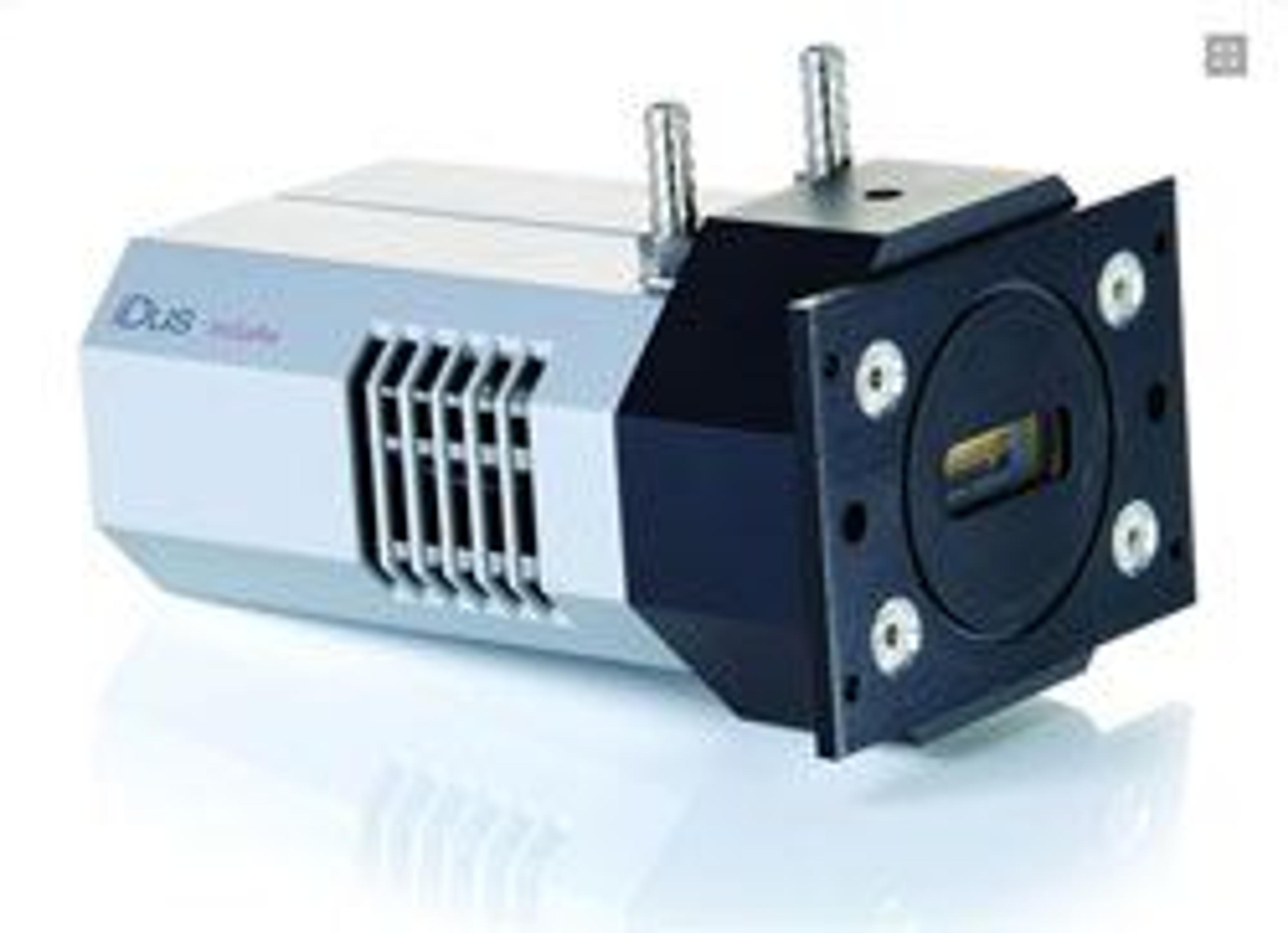
10 Oct 2011Nottingham University researchers demonstrate a non-invasive Raman microspectroscopy technique that delivers high purity cell populations. The team used an Andor iDus 401A-BRDD cooled, deep-depletion, back-illuminated CCD camera attached to a purpose-built Raman microspectrometer to record spectra from individual cells derived from micrometric regions of human embryonic stem cells (hESC).
Many medical researchers believe that stem cell therapy will revolutionise the treatment of human disease and may provide treatments for many currently incurable diseases. However, one problem still to be overcome is controlling the excessive proliferation of cells with unwanted phenotypes after transplantation to prevent tissue overgrowth and tumour formation.
Unfortunately, most techniques currently available for the characterisation of cells are invasive and make the cells unusable. Now, a Nottingham University team led by Dr Ioan Notingher has developed a non-invasive Raman microspectroscopy (RMS) technique that phenotypically identifies live cardiomyocyte cells within highly heterogeneous cell populations with greater than 96% sensitivity and specificity.
The team compared spectra recorded on the Andor iDus 401A-BRDD from individual cells derived from micrometric regions of hESC with matching immunofluorescence images from the same cells, they showed that the Raman spectra correspond to the spatial distribution of biomolecules such as nucleic acids, proteins, lipids and carbohydrates, and that this can be used to discriminate between different cell types.
“We needed the shortest possible acquisition times for the Raman spectra and the Andor iDus 401A camera is ideal for this application, allowing measurements of Raman spectra from selected positions in the cells in only 0.5 seconds,” says Dr Ioan Notingher. “Also, the detectors are optimised for the spectral regions in which we work, 800-900 nm, which is vital for avoiding photodamage to the cells. Since RMS has only a minimal background signal from water, it allows repeated observations of viable cells maintained under physiological conditions.
“Together with Professor Denning, also of the University of Nottingham and a leader in regenerative medicine, we have validated the potential of RMS for allowing the non-invasive phenotypic identification of hESC progeny. With further development, such label-free optical techniques may enable the separation of high-purity cell populations with mature phenotypes and provide repeated measurements to monitor time-dependent molecular changes in live hESCs during differentiation in vitro.”
According to Antoine Varagnat, Product Specialist at Andor, “the iDus has been the detector of choice in the research community for many years and Dr Notingher’s breakthrough work is a fantastic testimony to the performance of this platform. With its unique -100°C thermo-electric cooling platform and highest sensitivity in the near-infrared (NIR), as well as an extremely compact design for ease of integration to complex experiments, the iDus is just the right camera for NIR micro-Raman applications.”.
Compared to existing techniques, Raman microspectroscopy has several unique advantages for characterizing heterogeneous cell populations that makes it ideal for stem cell therapy. Conventional cell biology assays, such as PCR and Western Blotting, are invasive, require a large number of cells, and present averaged results representing entire cell populations. Fluorescence and magnetic cell sorting approaches rely on lineage-specific surface markers expressed on the cell membrane. However, many cell types, including cardiomyocytes, do not express these markers on the surface and are rendered unusable in a clinical environment by fixation and permeabilization. And, although transgenic strategies may be used to express surface markers, the complex genetic modifications also mean that the resulting cells are not suitable for clinical applications.

