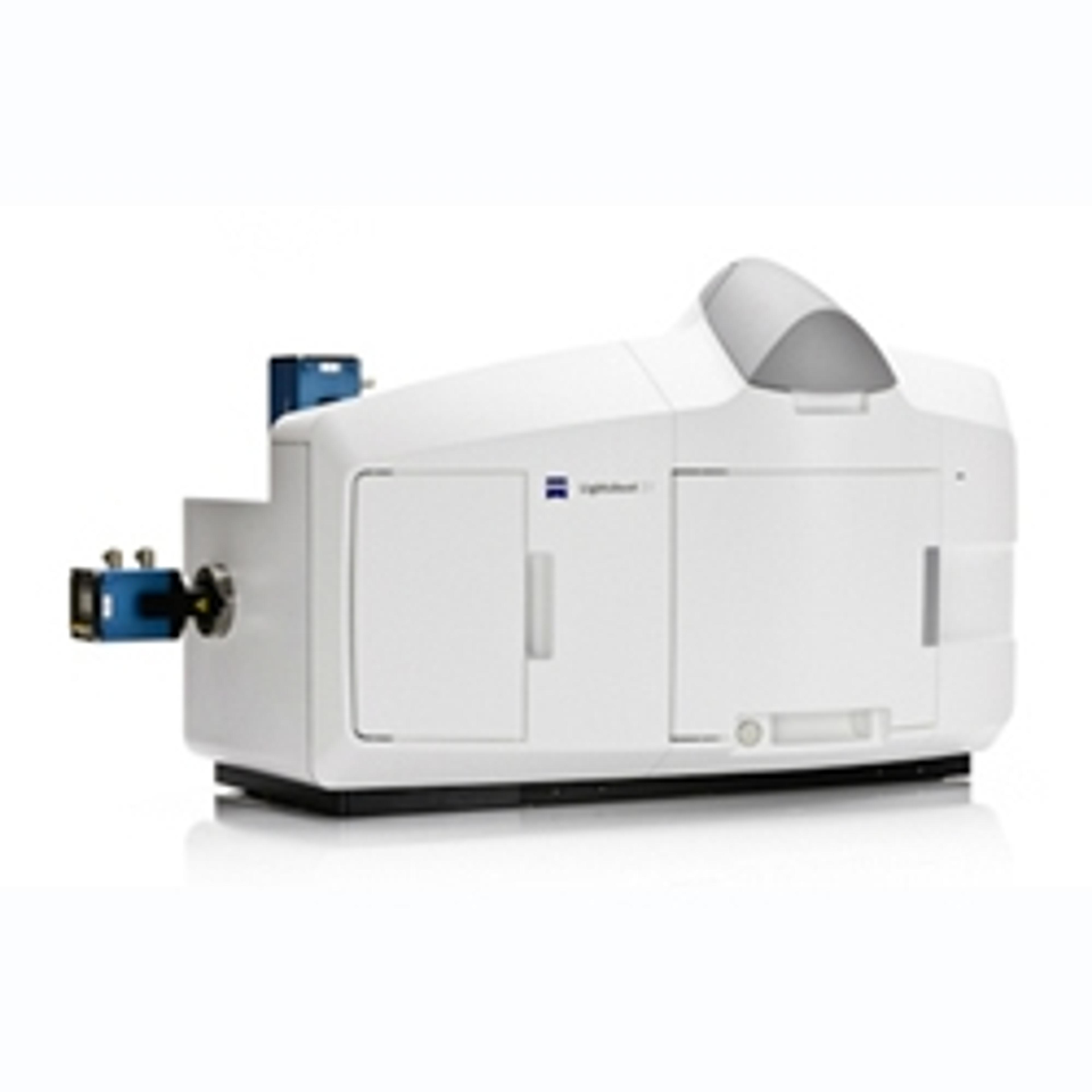
Carl Zeiss Introduces Lightsheet Z.1 LSFM Imaging System at Neuroscience 2012
14 Oct 2012The Microscopy business group at Carl Zeiss is presenting a new technology at the Society for Neuroscience Annual Meeting in New Orleans, Louisiana. The Lightsheet Z.1 provides biologists with a new method of imaging dynamic processes in living organisms.
Biologists can use the new microscopy system to observe the development of entire organisms over several days or more. The extremely low phototoxicity and the integrated incubation enable gentle imaging of specimens over hours to days. On large objects, in particular, such as fruit fly or zebra fish embryos, the light sheet microscope delivers more information than established methods of fluorescence microscopy. "The bigger the sample, the more you can get out of it with light sheet microscopy," says Dr. Pavel Tomančák from the Max Planck Institute for Molecular Cell Biology and Genetics in Dresden, Germany, describing the benefits of the new method. Lightsheet Z.1 can also be used in marine and cell biology, as well as plant physiology.
Lightsheet Z.1 works with an expanded light beam (the light sheet) that illuminates only a thin section of the sample, thus protecting the rest of the specimen. Images are captured at a 90 degree angle to the light sheet. Therefore, Lightsheet Z.1 achieves maximum image quality at minimal illumination intensity and is particularly well-suited for long-term examinations of living specimens. Multiview imaging allows data acquisition from different viewing angles. These can be combined through mathematical algorithms into 3D reconstructions and time-lapse videos.
The light sheet system of the Lightsheet Z.1 uses a new type of optical concept that combines cylindrical lens optics with laser scanning. Users receive homogeneously illuminated optical sections of complete examination objects.
Dr. Olaf Selchow, Product Manager for Light Sheet Microscopy at Carl Zeiss states: "I'm confident that this illumination principle is going to revolutionize 3D fluorescence imaging."

