Chemistry Nobel Prize 2014: Super-Resolution Fluorescence Microscopy
31 Oct 2014
SelectScience would like to congratulate Eric Betzig, Stefan W. Hell and William E. Moerner on winning the 2014 Chemistry Nobel Prize 'for the development of super-resolved fluorescence microscopy'.1
Previously, confocal microscopy was used by many scientists to view living systems. However, by using a single beam of light it was difficult to focus on a specific area, and the light has the detrimental effect of degrading the sample over time. Recently, the Nobel Prize winners have developed a new way of viewing living systems using light microscopy, enabling scientists to view living tissues down to a molecular level (~20nm) with the resolution exceeding the diffraction limit.
PALM Microscopy
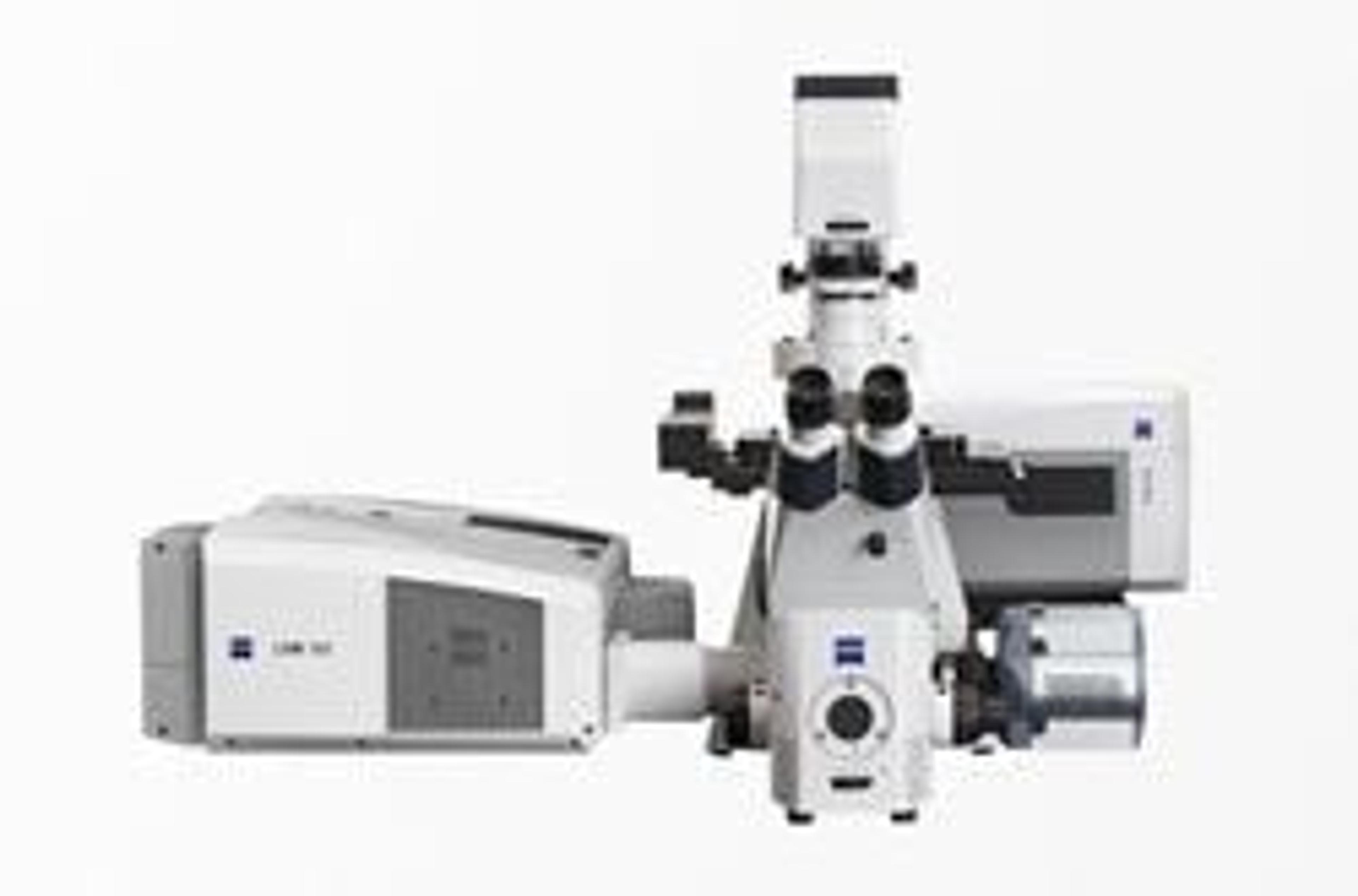
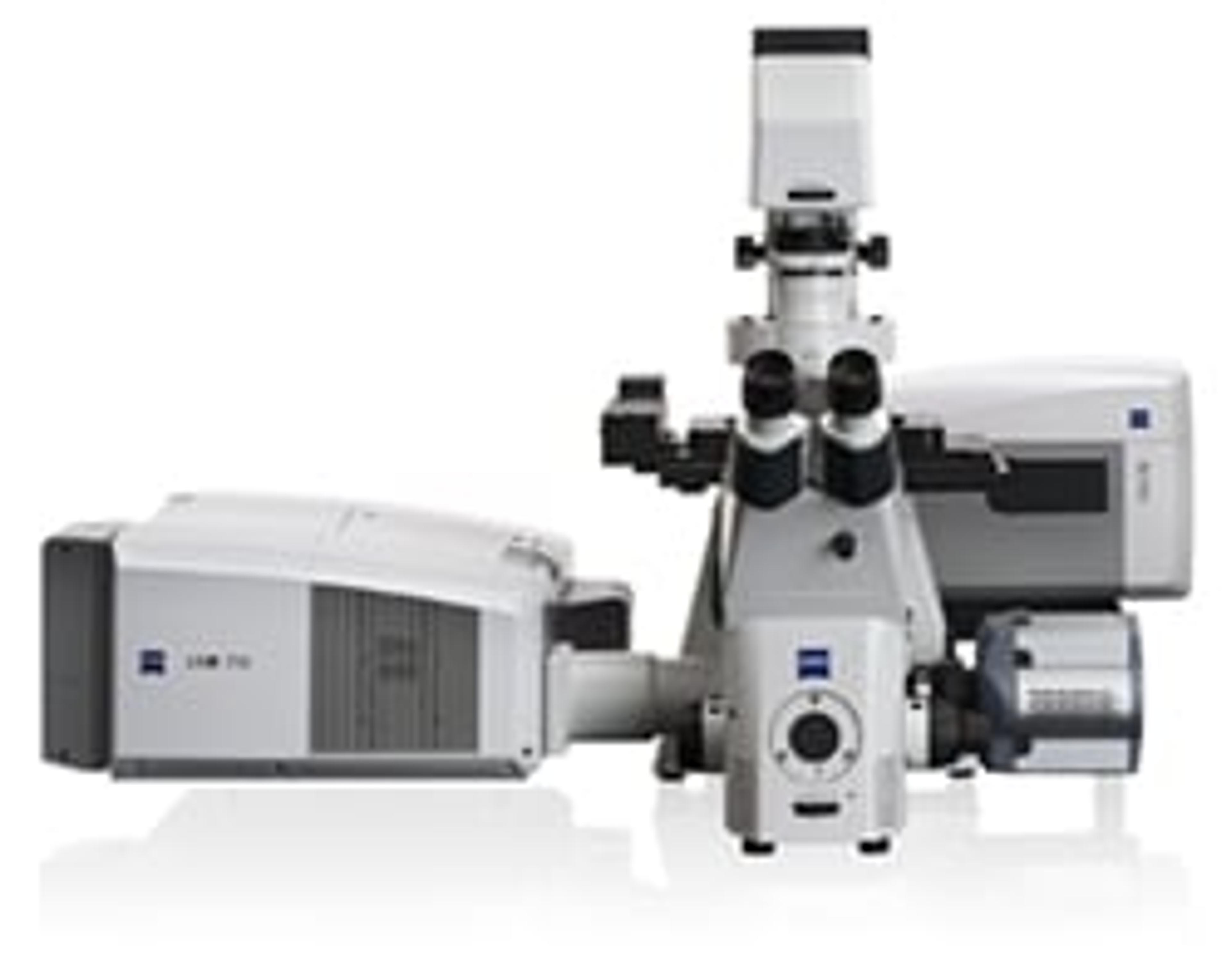
Eric Betzig of Howard Hughes Medical Institute's Janelia Research Campus, together with ZEISS, has developed the world's first commercially available 3D super-resolution fluorescence microscope. The ZEISSELYRA Super-Resolution Microscopy Systems are available in two forms;
ELYRA S.1 uses super-resolution structured illumination microscopy (SR-SIM) that enables scientists to view in fine structural details samples labeled with conventional dyes.
ELYRA P.1 uses the Nobel Prize winning photo-activated localization microscopy (PALM) that enables scientists to view endogenously-expressed photo-switchable fluorescent proteins by localizing small molecules with a resolution down to 20nm laterally and 50 nm axially.
Since winning the Nobel Prize for Chemistry, Professor Betzig has gone on to develop a new microscopy technique that enables imaging of fast-moving living systems using lattice sheets of light instead of cones.
STED Microscopy
Stefan W. Hell of Max Planck Institute, together with Leica Microsystems, has developed the world's first stimulated emission depletion (STED) microscope. The Leica TCS SP8 STED 3x enables scientists to study sub cellular architectures with nano-scale resolution.
Super-resolved fluorescence microscopy has revolutionized the way scientists can view living cells. The ability to monitor the division and growth of living cells gives in insight into the way in which cancer grows, viruses duplicate and the effect of neuro-diseases on brain tissue.
Ref: 1) http://www.nobelprize.org/nobel_prizes/chemistry/laureates/2014/ Accessed 31.10.14
Learn more about microscopy: 2014 SelectScience Microscopy Buying Guide