CLINICAL24: How AI is driving the future of flow cytometry and regenerative medicine
Dr. Vasiliki Kalodimou, CLINICAL24 Ambassador, shares how her team is advancing regenerative medicine while embracing new technologies to accelerate discoveries.
17 Oct 2025
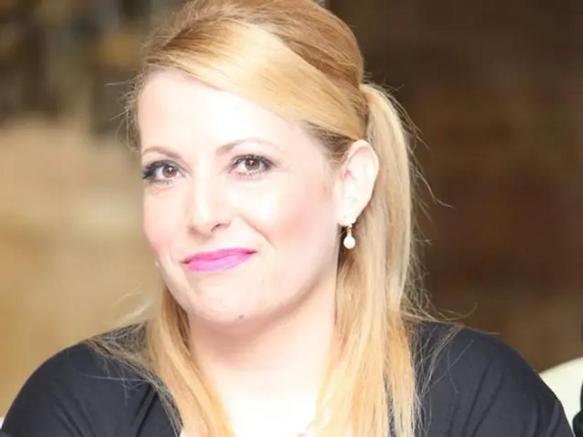
Dr. Vasiliki E. Kalodimou, Director of the Flow Cytometry–Research and Regenerative Medicine Department at EUC Frankfurt Branch
Dr. Vasiliki E. Kalodimou, now in her second year as a CLINICAL24 Ambassador, and Director of the Flow Cytometry–Research and Regenerative Medicine Department at EUC Frankfurt Branch, shares how her team is advancing regenerative medicine while leveraging AI to accelerate discoveries. In this interview, she reflects on her lab’s progress over the past year, from high-parameter flow cytometry to stem cell-based regenerative applications, and highlights how artificial intelligence is transforming workflows and driving clinically actionable insights in both research and patient care.
Advances in cytometry and regenerative medicine
“It’s been a really exciting and productive year since CLINICAL24,” says Dr. Kalodimou. Her lab has been driving advances on several fronts. In flow cytometry, the team has expanded into high-parameter and spectral panels, pushing them to refine panel design and improve standardization of large datasets. At the same time, AI-assisted gating and clustering tools are being explored, bringing greater efficiency and reproducibility to workflows.
Beyond cytometry, the lab has focused on mesenchymal stem cell (MSC) research and clinical trials in orthopedics. By characterizing, optimizing, and monitoring MSCs, the team aims to unlock their regenerative potential. Exosomes, a key component of the MSC secretome, are also under investigation as both biomarkers and therapeutic tools for orthopedics and beyond.
In parallel, Dr. Kalodimou’s team has begun laying the groundwork for 3D organ printing. “We’ve been focusing on building the right infrastructure and workflows to help these technologies move from experimental models toward translational and clinical applications,” she explains.
Flow cytometry as a multimodal, AI-driven powerhouse
Looking ahead, Dr. Kalodimou sees flow cytometry evolving into a multimodal, AI-powered analytics powerhouse. AI-enabled gating and automated analysis are already accelerating workflows, delivering reproducibility, speed, and the ability to detect rare or subtle populations. Tools such as GateNet and FlowJo integrations are helping labs standardize pipelines, with regulatory-grade AI solutions for clinical use on the horizon.
3D imaging and tomographic cytometry are adding a new dimension to cellular analysis. Imaging flow cytometry is moving beyond 2D snapshots to high-throughput 3D reconstructions, capturing subcellular morphology and enabling morphology-aware sorting. Paired with AI, these approaches allow researchers to detect rare events and discover novel biomarkers while reducing reliance on large antibody panels.
Microfluidic and point-of-care cytometers are also democratizing access to cytometry. Compact, AI-enabled devices can now deliver accurate counts and phenotyping from whole blood with minimal preparation, shortening time-to-result and bringing advanced analysis to low-resource settings.
Meanwhile, spectral and mass cytometry continue to expand panel depth, with 30–40 marker panels now routine and CyTOF enabling ultra-high-plex profiling in oncology and immunotherapy. Advances in fluorescent dyes and nanobody reagents further improve sensitivity and reduce spectral overlap, supporting the detection of rare cell populations.
“These advances are transforming cytometry from a fluorescence-based phenotyping tool into a multimodal, spatially-resolved, and clinically actionable platform,” says Dr. Kalodimou. As these technologies mature, they promise greater accessibility, precision, and insight, ushering in a new era of single-cell and tissue-level analysis.
AI and machine learning as key drivers
Artificial intelligence and machine learning are already reshaping workflows in Dr. Kalodimou’s lab.AI-assisted gating tools have drastically reduced analysis times, cutting hours of manual work down to minutes while producing reproducible population maps. Clustering algorithms such as FlowSOM and UMAP help make sense of increasingly complex, high-dimensional data, enabling the exploration of immune subsets without oversimplification.
“AI and ML are not just ‘add-ons,’ they are becoming central to how cytometry data is collected, processed, and interpreted,” she explains. This shift is evident across multiple fronts: AI enhances sensitivity for rare cell populations, uncovering subtle, biologically meaningful subsets that might otherwise be overlooked. Combining imaging flow cytometry with machine learning allows cell shape and subcellular features to be incorporated alongside fluorescence data, opening new avenues for experimental questions.
Beyond analysis, AI is improving consistency across experiments and operators, a critical factor for multi-site collaborations and clinical studies. Challenges remain, including training the team to use ML tools effectively, managing large imaging and spectral datasets, and interpreting model outputs to understand how populations are classified.
“The future is AI-driven cytometry — faster, standardized, multimodal, and clinically actionable,” says Dr. Kalodimou. “AI isn’t replacing human expertise, but it is accelerating routine tasks and revealing patterns that guide our next hypotheses.”
What labs need to embrace new technologies
For these technologies to take root in day-to-day practice, Dr. Kalodimou highlights the importance of hands-on training, approachable AI education, strong technical support, and institutional investment.
“In my lab, what really makes it easier to adopt new tools and technologies is support that’s practical and hands-on,” she explains. Short, workflow-focused training sessions, she says, are far more useful than long theoretical ones, particularly when they involve real datasets that mirror experiments.
She adds that data analysis and AI skills are becoming increasingly essential: “As panels get more complex and imaging data grows, we need approachable ways to understand machine learning outputs and integrate them into our analysis without becoming data scientists ourselves.”
Ongoing technical support, whether from application specialists or user communities, also helps ensure smoother adoption. At the institutional level, investment in data storage, computing infrastructure, and dedicated training time are seen as key enablers.
Looking ahead, Dr. Kalodimou believes that lowering these barriers will be critical to unlocking the full potential of AI-driven cytometry and regenerative medicine, making advanced technologies practical and accessible so researchers can focus on biology rather than bottlenecks.