Colony and organoid formation: accelerating cancer drug discovery
26 Nov 2024The colony formation assay (CFA), also known as the tumour forming assay, is a relatively simple yet ubiquitous and powerful tool in cancer research thanks to its ability to evaluate the long-term proliferative potential of cancer cells. The assay offers unique insights into the capacity of individual cells to survive, grow, and form colonies, reflecting a fundamental aspect of cancer cells: their ability to proliferate indefinitely and form tumours.
The CFA has become a gold standard technique with demonstrated effectiveness in the following key research areas:
- Assessment of tumorigenicity
- Testing therapeutic agents
- Study of cancer cell invasiveness and resistance
- Investigation of genetic and molecular pathways
- Cancer progression and metastasis research
The CFA continues to contribute to our understanding of cancer and therefore to accelerating the development of effective cancer treatments. Meanwhile the growth of 3-dimensional organoids, derived from a variety of primary cell types has revolutionized the study of disease and therapeutic responses in vitro. Organoids consist of 3-dimensional multicellular structures that resemble and mimic in vivo tissue architecture and function.
The rate limiting step
While the colony formation assay is inherently time-consuming, typically requiring 1-3 weeks of cell growth in an incubator, the rate-limiting, labour intensive and most subjective stage is the subsequent scoring and analysis of colonies generated.
Similar to the CFA, organoid growth assays also rely on accurate object scoring and, especially, accurate organoid size determination. This has traditionally been a manual process, often tediously performed under a microscope. Not only is this approach subject to operator bias and fatigue, but it also offers no practical path to determining colony, spheroid, or organoid size.
In the case of the CFA this is a serious shortcoming as cancer treatment regimes may in many cases not affect the number of colonies generated but instead affect the rate of colony growth and thus average colony size. A nuance that risks being missed altogether with manual processing of the CFA.
The GelCount™ advantage
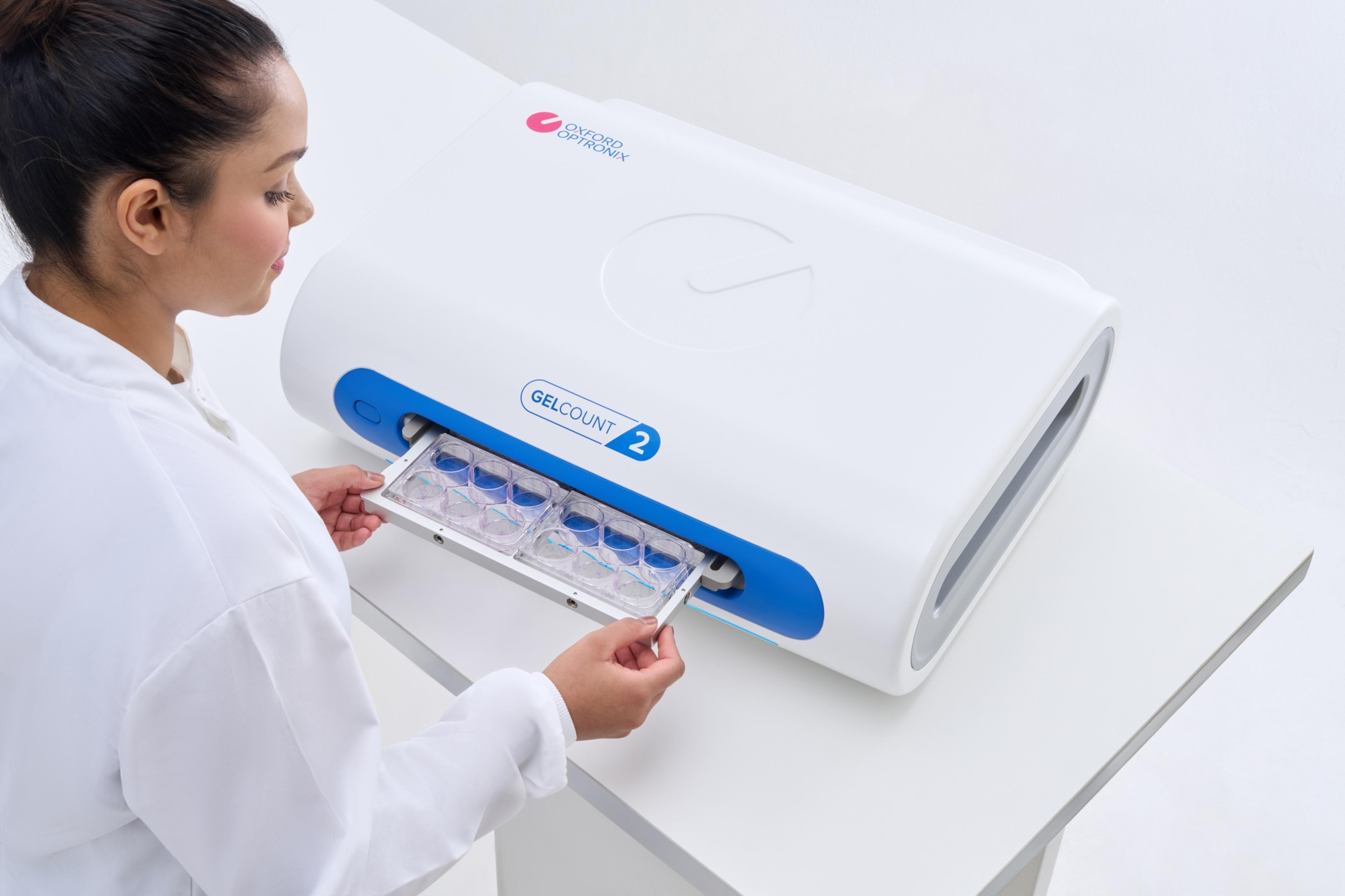
The solution is to digitize the process. The GelCount™ is a high-resolution imager capable of generating high-quality images of colonies, spheroids and organoids cultured in multi-well plates and Petri dishes. GelCount™ is an all-in-one solution that not only generates images but automatically processes them. Output includes both a numerical count of object numbers, and crucially, a full breakdown of object diameter in um.
Over recent years the GelCount™ platform has become the go-to imaging device for cancer biologists and life scientists around the globe seeking to accelerate processing of in vitro colony, spheroid and organoid formation assays. And for 2025, Oxford Optronix is proud to build on this legacy by unveiling GelCount™ 2, a second-generation implementation featuring fully redesigned hardware for faster imaging, and a powerful new version 2 software platform to match.
References
- Strand Z. et al. 2023 (Cancers) Establishment of a 3D model to characterize the radioresponse of patient-derived glioblastoma cells.
- Urban-Wójciuk Z. et al. 2022 (Cancer Gene Therapy) The biguanide polyamine analog verlindamycin promotes differentiation in neuroblastoma via induction of antizyme.
- Hatch S. et al. 2022 Automated 96-well format high throughput colony formation assay for siRNA library screen.

