CT Scanner Facility at Stellenbosch University in South Africa Applies Deben tensile stages in X-ray CT Analysis and 3D Printing Projects
5 Jan 2016
Deben, a leading provider of in-situ testing stages together with innovative accessories and components for electron microscopy, has supplied various tensile stages for use in X-ray computerized tomography (CT) analysis to the CT Scanner Facility at Stellenbosch University in South Africa.
Dr Anton du Plessis is Manager of the CT Scanner Facility at Stellenbosch University in South Africa. The Facility acts as a CT Scan Service provider with the aim to provide a high quality 3D imaging and analysis service, with fast turnaround times. The unit serves clients from both academia and industry from all over South Africa as well as many from overseas.
Dr du Plessis talks about the choice of stages and how they are being used: “We decided to purchase the Deben in situ stages to add value especially in applied research projects from the engineering users of the facility. The acquisition was not for one specific project as ours is a multi-user facility focusing on providing top quality and unique facilities for researchers and industry to make use of. In the first year since installation, we have looked at some interesting samples using the in situ stages. These have included compression tests on wood samples and then visualizing the cell wall damage that occurs during compression as one nice example. Some tests have been done on glass and carbon fiber composites where the interest is to test different types of layering patterns and to study and optimize the strength properties of these composites. There is also an in-house research interest in 3D printing and additive manufacturing (the metal form of 3D printing). It is quite clear that the new Deben in situ X-ray CT tensile and compression stages will be applied in this work too and we are very much looking forward to that. In the short term we have already 3D printed some ABS plastic samples and started testing them and plan on visualizing the exact internal point of failure.”
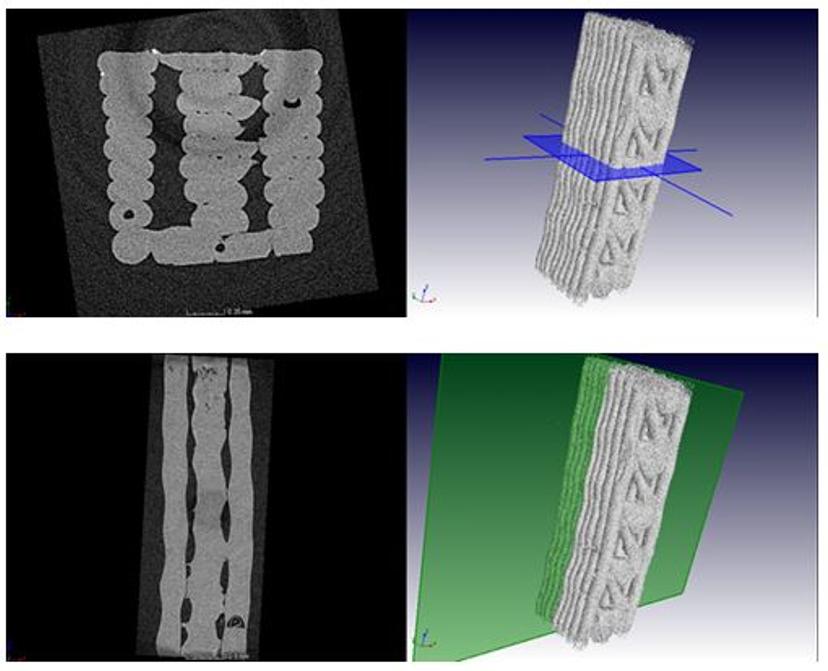
3D CT scans at 40 N show the internal structure of the central section of the tensile sample. The brighter grey is plastic and black is air. The 3D view with plane shows the orientation of the 2D slice view to the left.
“One of the major advantages of CT imaging is the ability to non-destructively visualize materials before and after processing (e.g. compression or pulling), therefore having an in situ stage allows researchers to visualize the microstructural details inside their samples under load for the first time. Having it available as a core facility provides many more researchers access to this kind of testing which has formerly been limited to some synchrotrons and a few selected CT facilities worldwide. Our Stellenbosch CT Facility is fully open access so we hope this leads to some great new materials science discoveries.”