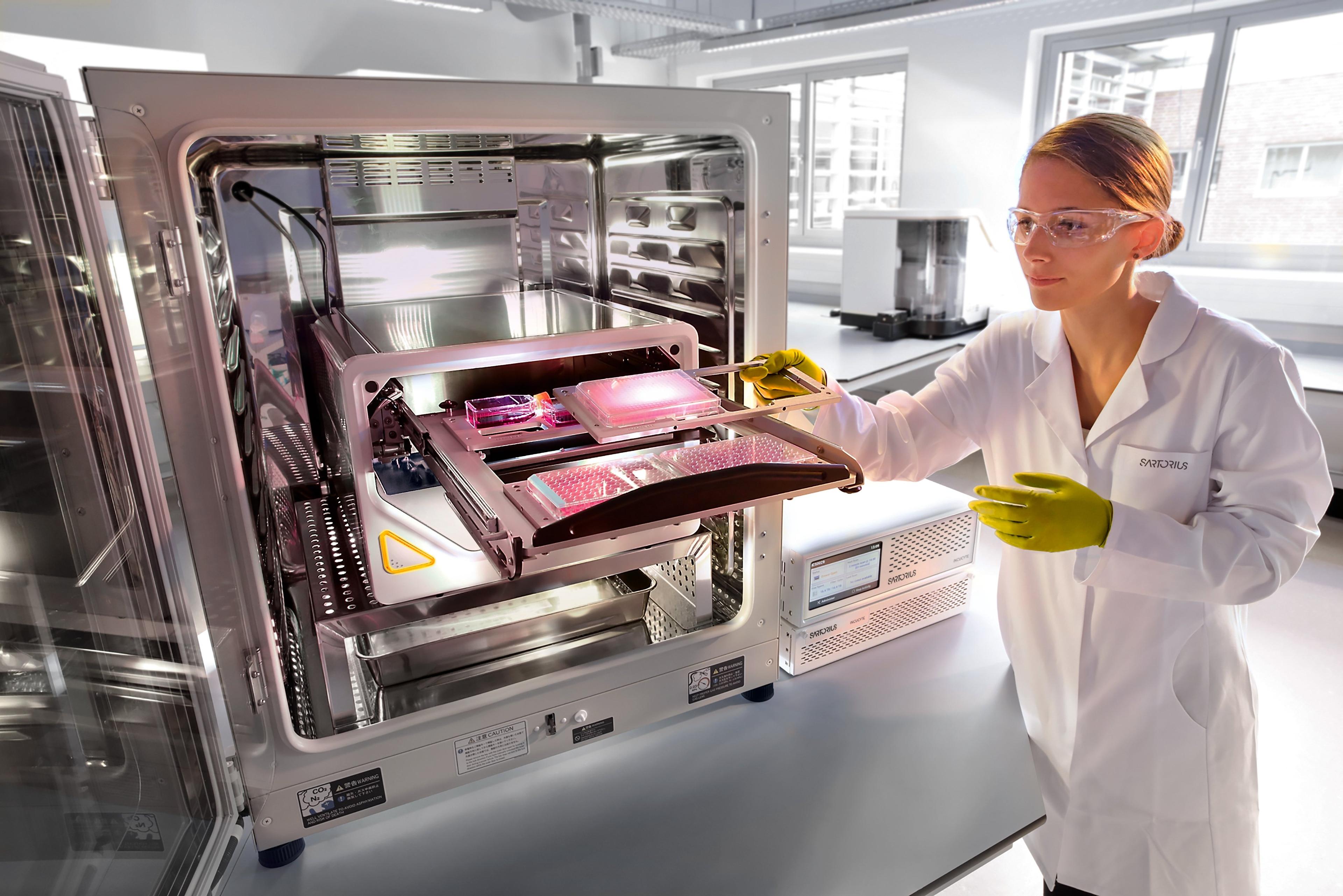
Evaluation of checkpoint inhibitor therapies using a mixed lymphocyte reaction (MLR) assay
Watch this on-demand webinar to learn how the iQue® Advanced Flow Cytometry Platform and associated reagent kits can be used to quantify T cell response in MLR
20 Sept 2021
Immune checkpoints are molecules that signal and control the synapse between T cells and antigen presenting cells (APCs). Checkpoint inhibitor therapies aim to target and inhibit the signals that ‘switch off’ T cells, leading to upregulation of T cell activation and tumor cell killing.
The mixed lymphocyte reaction (MLR) mimics the T cell and APC synapse by co-culturing immune cells from two individuals. Consequently, MLR is used as a model, to discover the effects of checkpoint inhibitor drugs in vitro, furthering biological research applications and drug discovery processes.
In this on-demand SelectScience® webinar, Kirsty McBain, Associate Scientist, iQue® Applications Group, Sartorius, outlines:
• The use of the iQue® MLR assay to generate pharmacological outputs for checkpoint inhibitor induced T cell activation
• The impact of checkpoint inhibitor therapeutics on inflammatory cytokine release
• How the iQue® Advanced Flow Cytometry Platform and associated reagent kits can be used to quantify T cell response in MLR
Read on for the live Q&A session or register to watch the webinar at a time that suits you.
Q: What are the advantages or disadvantages of using one-way compared to two-way MLR assays?
KM: The advantage of the one-way model is that you get a simple and unidirectional activation of T cells without any background signal, which is probably the main disadvantage of the two-way model. This is a bit more of a crude measurement of activation in cytokine release with a lot of increased background. The disadvantage of the one-way MLR is that it requires isolated immune cell populations. So this is a bit more time-consuming to prepare and more expensive to acquire, compared to just the simple peripheral blood mononuclear cells (PBMCs) that are required for the two-way MLR.
Q: For the two-way MLR, can you label in a way that allows proliferation of T cells from each donor to be distinguished?
NB: Yes, there are three different configurations of the Encoding Dye Kit in the iQue® portfolio. There is the violet-blue, the blue-green, and the red-red. The reason we've only used the blue-green design in the experiment is because it's compatible with the markers in the T Cell Activation Kit. But, if you want to use the multiple proliferation design, then these can still be analyzed alongside cytokines and/or when you design an antibody panel.
Q: How does this assay compare to traditional MLR assays?
KM: The more traditional techniques for measuring T cell response in MLR generally require separate assays to be carried out for the marker expression and cytokine release, for example, a more traditional flow cytometry, alongside another assay such as an ELISA. The main benefit here of the iQue® workflow is that we can measure all of these parameters in a single well. So it really speeds up the time for both the assay and the analysis. It also reduces the amount of the sample you're going to need and reduces the chances of there being variation due to measuring different outputs from different cell populations.
Q: What are the other uses of MLR?
KM: Prior to the use of MLR we've shown here, which is to look at the effects of checkpoint inhibitors, the more traditional use of MLR is to check the safety of a drug or therapy. For example, it could be used as an initial way to check whether an allogeneic cell therapy is going to be likely to result in immune rejection.
Q: For multi-day cytokine measurements, given the suggested one-time use of the provided standards, are you freezing supernatants and running them all at once or using the standards on multiple days?
KM: Yes, we tend to freeze the cytokines and then analyze at the end. So we just take 10-microliter samples, transfer to V bottom plates, freeze those, and then add cytokine quantitation directly to those frozen plates. It means we can use the same standards across all the different time points.
Q: Are your DCs from frozen stock or do you differentiate from monocytes and place them straight into culture?
KM: Just for ease, we have always used frozen DCs rather than differentiating, but I'm sure you can do either/or.
Q: Can you explain how you quantify T cell proliferation in these MLR assays?
KM: Yes, the T cells are labeled before being added to the plate with a proliferation dye which comes with the T cell kit. This monitors the proliferation of the T cells over time, meaning that by the time we run on the iQue®, we can see how much the T cells have proliferated.
SelectScience runs 10+ webinars each month, discover more of our upcoming webinars>>