Expanding the immunohistochemistry (IHC) workflow with patient-free positive controls
In this guest article by RayBiotech Life, learn how to easily obtain IHC positive controls for different pathologies without the need for patient samples
13 May 2021
Immunohistochemistry (IHC), or the staining of cells or tissues for a specific antigen-of-interest (AOI), is a valuable tool in clinical diagnosis. Positive control samples are an essential quality control measure to properly assess affinity reagents and the IHC workflow. However, obtaining positive control patient samples for IHC can be difficult, particularly for infectious and rare diseases.
To address this issue, RayBiotech offers a service to develop patient-free positive IHC controls. This custom service inserts exogenous DNA encoding the AOI into human cells where the AOI is overexpressed. The cells are then pelleted and fixed with formalin, thus enabling the downstream staining of pellet sections for the AOI. Approximately 200 sections can be obtained with 1 standard cell pellet containing 90 million cells.
Service features:
- Customizable: Choose the target protein you want
- Formalin-fixed human cells
- Cells can be handled at biosafety level 1 (BSL1)
- Negative controls available
- Affordable
Handling COVID-19 samples safely
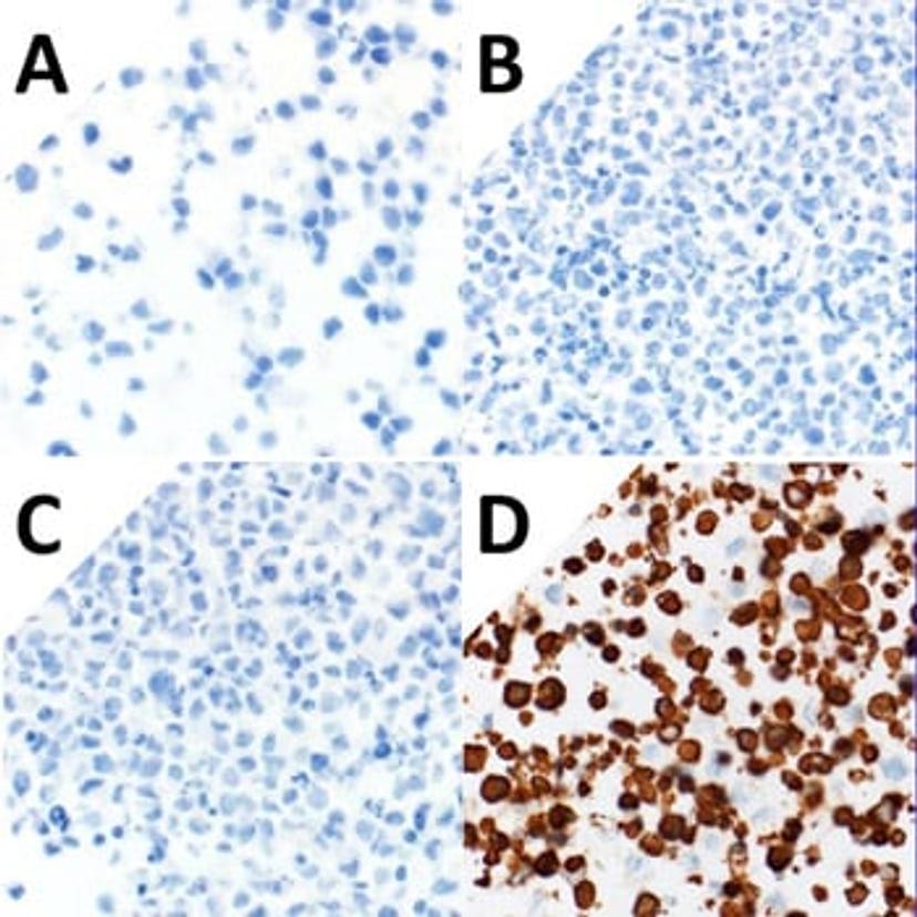
For one laboratory, obtaining and safely processing tissue samples from known COVID-19 patients proved challenging. RayBiotech prepared three patient-free IHC positive controls for the detection of different antigens produced by the severe acute respiratory coronavirus 2 (SARS-CoV-2). These antigens included the SARS-CoV-2 Nucleocapsid, Spike subunit 1, and Spike subunit 2 proteins. The IHC positive controls and a negative IHC control lacking exogenous DNA were then probed with an antibody to Spike subunit 2 (Figure 1). Only the cells overexpressing the Spike subunit 2 protein were stained.
Read more about the custom IHC control service>>
References
Szabolcs M, et al. Identification of Immunohistochemical Reagents for In Situ Protein Expression Analysis of Coronavirus-associated Changes in Human Tissues. Appl Immunohistochem Mol Morphol. 2021