Harnessing the Power of 3D Pharmacology for Improved In Vitro to In Vivo Predictions
Find out how BIOENSIS uses its novel 3D drug testing system to predict in vivo pharmacological reactions
20 Jun 2017

BIOENSIS is a pre-clinical contract research organization (CRO) that specializes in the pharmacology of spheroids. The company’s novel 3D drug testing system enables more accurate in vitro to in vivo predictions, preventing costly expenditures in downstream development. SelectScience® speaks to Gonzalo Castillo, Vice President of R&D and Business Development at BIOENSIS, to find out more.
SS: Can you give an overview of your work at BIOENSIS and a bit of background about the company?
GC: We are a company that focuses on 3D pharmacology. Our aim is to harness the power of 3D-cellular models, to provide better, more predictive pharmacology than the current in vitro methods. We utilize the power of 3D in vitro for both on-target and off-target applications.
For on-target activities, the focus of our services has been mainly in oncology. We have a broad panel of 3D-cellular models; tumor-derived cell lines that are routinely tested for our clients. We also provide invasion and migration assays, combination assays, as well as custom assay development. For off-target activities, we are developing a suite of organotypic models; mostly focused on toxicology.
Replicating in vivo models
SS: What are the advantages of 3D cell culture in pre-clinical development?
GC: Studies carried out in cell monolayers have limited translational value because they do not capture the known complexity of an organ or tumor microenvironment. 3D-cell cultures, on the other hand, account for cell-cell interactions, matrices, and formations of metabolic gradients, vital to understanding drug efficacy and resistance. 3D-tissue culture models capture the microenvironment and are one step closer to representing in vivo tumor responses, allowing for greater predictive power in drug candidacy.
SS: What projects are you working on currently?
GC: We work for pharmaceutical and biotech companies that want to test the pharmacology of their compounds in 3D-cell cultures. The focus of what we do is divided into two areas. For oncology, we have developed on-target models – our current platform has 94 human tumor models, and we use them to carry out testing for pharmaceutical companies. These companies are interested in discovering how a tumor model responds to a particular compound. Or, they want to associate biomarker sensitivity to a compound of interest. Pharmaceutical companies use the information that we provide to make more informed decisions about whether to move a compound into in vivo models or clinical trials.
We are also developing off-target organotypic models, to replicate the organs you would find in the body, for the testing of skin, kidney and liver toxicities.
SS: How do the Elplasia plates of Kuraray enable your work and why did you choose to use these particular plates?
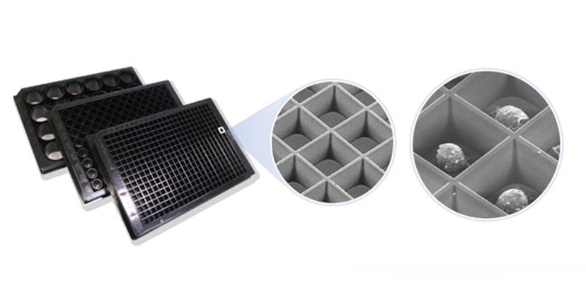
GC: The Elplasia plates of Kuraray are incredibly well engineered to pharmaceutical drug research: for many different reasons. One of the most important considerations, when you are testing compounds, is that you are trying to determine which compound is most efficacious. The plates we use must be highly reproducible, the model must be relevant, they must be automatable, and they have to show minimum variation between data points. The ELPLASIA™ plates meet these objectives.

One unique aspect of the Elplasia™ plates (and what differentiates them from other available plates) is that there are multiple spheroids per well and the spheroids are a very reproducible size ideal for higher throughput. We use the Elplasia plates extensively because they allow for one step scaffold-free formation and analysis of spheroid pharmacology, the data we obtain utilizing these plates are again very reproducible, automatable and they are very flexible allowing multiple detection modes especially automated imaging.
SS: What challenges do you face in developing customized assays and 3D cell cultures, and how do you overcome these?
GC: 3D-cellular models are complex. Forming accurate spheroid models involves determining the optimal microenvironment that will allow for the spheroid to form and survive for the time of the experiment. You may also need to account for other cellular components; for example, if you need to reconstruct the heterogeneity of a tumor. This fine tuning makes every single model a challenge!
We often customize assays for our clients, and this can also be quite challenging. All the components that go into the media of the 3D-cellular model have to be strictly controlled and validated. But; while customization can be complex, the resulting model will be far more predictive of an in vivo outcome.
SS: What are your plans for the future direction of BIOENSIS?
GC: Historically, we have focused heavily on oncology and will continue developing models to support this therapeutic area. The area we have identified for future development is in toxicology, and are actively utilizing our expertise to develop organotypic models for skin, liver, and kidney. The Elplasia plates of Kuraray are ideally suited for this application, and we will continue to use them as we move forward with these new platforms.
