High Resolution 3D Light and Electron Microscopy Analysis of Living Tissue at the University of Ghent
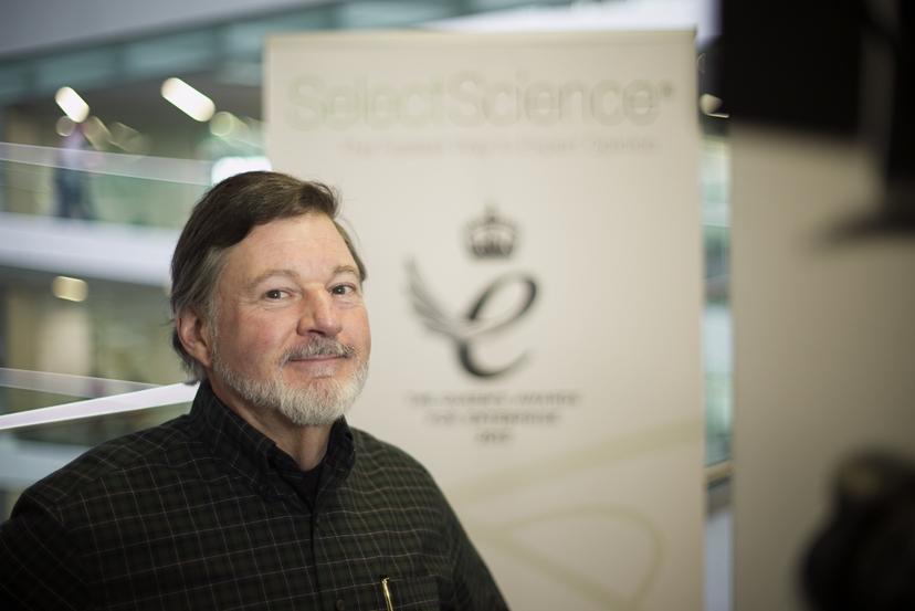
SelectScience® spoke to Chris Guerin, Bio Imaging Core facility at the Flanders Institute of Technology, about innovations in high resolution correlative microscopy
31 May 2016

SelectScience® spoke to Chris Guerin, Bio Imaging Core facility at the Flanders Institute of Technology, about innovations in high resolution correlative microscopy
European Molecular Biology Laboratory (EMBL) Founded in 1974, EMBL is Europe’s flagship laboratory for the life sciences – an intergovernmental organization with more than 80 independent research groups covering the spectrum of molecular biology.
At the Flanders Institute of Biotechnology (VIB), Christopher Guérin leads the Ghent Bio Imaging Core. This core facility is developing advancements to 3D Correlative Light and Electron Microscopy, serving not only over 1200 scientists at the VIB, but members of the scientific community all over the world. At the workshop ‘From 3D Light to 3D Electron Microscopy’ (EMBL 3D), jointly organized by the European Molecular Biology Laboratory (EMBL) and ZEISS Microscopy, SelectScience spoke to Chris about the innovations coming out of this site.
When the core facility was originally being set up, “the managing director came to me and said ‘what do we need to make it as good as anything in the world?’ and I said “we need one of everything”, Chris explained. “In fact that was the wrong answer- we need several of most things.” The Ghent facility specializes in volume electron microscopy, which is “electron microscopy in three dimensions”. Using “high end instrumentation” such as confocal microscopes, super-resolution microscopes or electron microscopes, researchers at VIB can look at “the smallest details of a cell or a tissue in three dimensions at whatever resolution scale that we want”, from nanometers, to angstroms.
Doing microscopy at very high resolutions is particularly important in biomedical research “where ideally, we want to look at living things – we want to look at cells and tissues and how they function normally in healthy people”. It is also very important to understand “what happens when there is a mutation or when tissue is injured, how it either tries to repair itself or how it eventually fails and dies”, explained Chris. While it’s possible to look at “small things” with a light microscope, an electron microscope is required for “the fine structures of the cells, the smallest details”, Chris continued.
Imaging at the highest resolution
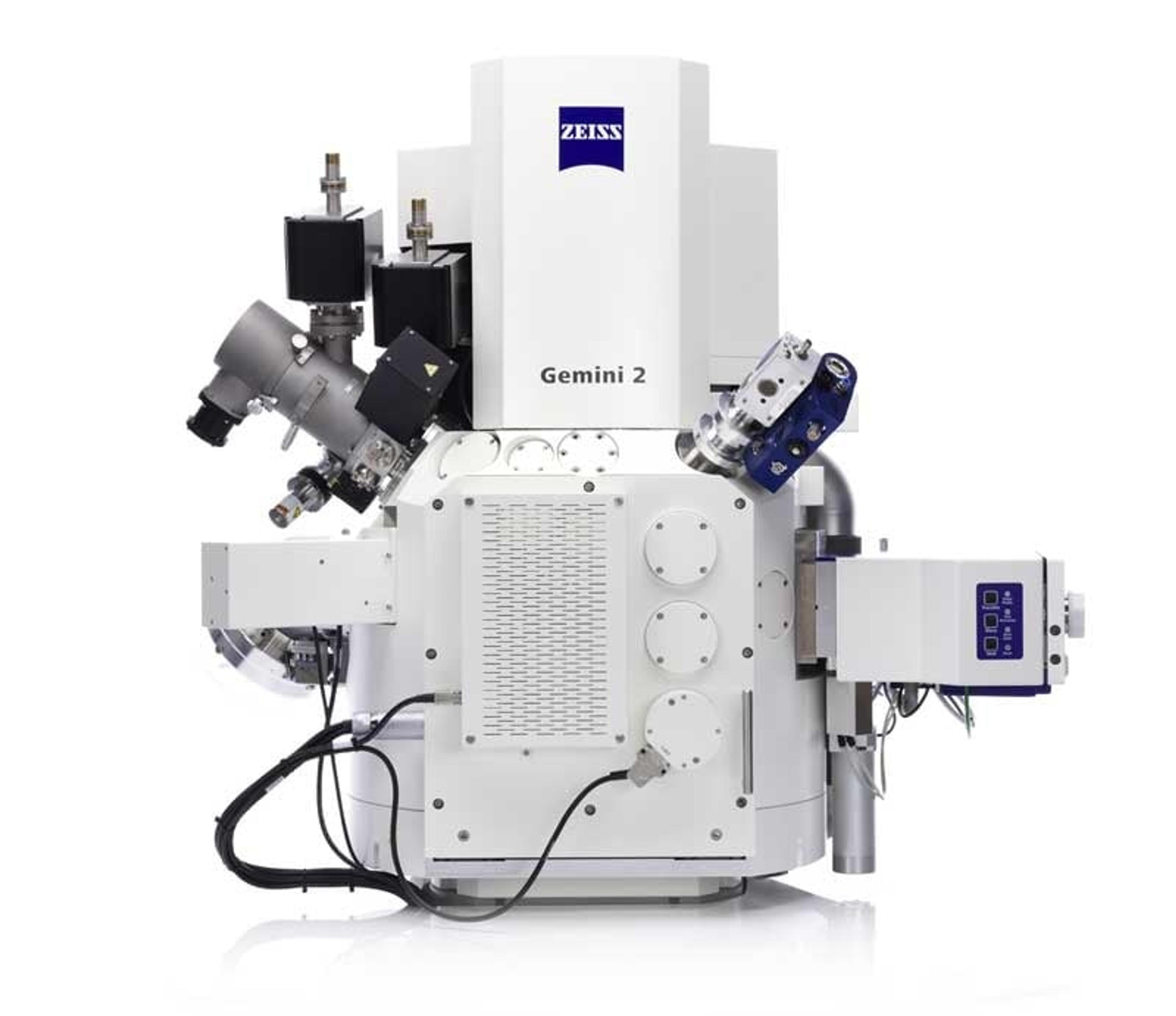
Two “very special microscopes” allow imaging in these high resolutions. At Ghent, “we have a ZEISS MERLIN that has a Gatan 3View on it”, a robotic ultramicrotome that allows 3D imaging of cells and tissues by forming a composite image from images of multiple thin slices. “As if you have a loaf of bread that was your cell, you slice that off, look at the first slice then, you slice again. When you get to the bottom crust, you can take all these slices and put them back together digitally so that you can see the entire tissue in three dimensions”. While the Gatan 3View “slices the bread in relatively thin slices”, for “really thin slices, with the highest details, we’re using the AURIGA or the Crossbeam 540, which allows us to get a very, very fine detail in both X, Y and Z”. Once these images are rebuilt, “we can really look at the basic building blocks of cells”.
EMBL3D
This is the third conference on correlative microscopy, “which is a small community but growing rapidly”, Chris explained. “These conferences are very important because they allow us to get together and share, which is a very critical thing to the advancement of this technique and the spreading of this technique throughout the scientific community.” Because individual laboratories may not have the expertise to implement this correlative technique themselves, core facilities and partnerships with sponsors like ZEISS “is very, very important”.
Find out more about the other speakers and presentations from EMBL 3D, or learn more about the applications of Correlative Microscopy.
Watch our interview with Chris here.


