High-resolution imaging leads the way in forensic pathology
Learn how one forensic pathologist has adopted high-resolution imaging technology to visualize forensic evidence with exceptional clarity.
14 Aug 2024
Dr. Marianne Hamel, a board-certified forensic pathologist practicing in Eastern Pennsylvania and New Jersey, discusses the impact of advanced high-resolution imaging technology on forensic pathology investigations, with a keen focus on the DP75 digital microscope camera from Evident.
Forensic examinations require meticulous methodology and precision. In this field, where each piece of evidence holds the potential to unravel mysteries and dispense justice, the role of high-resolution imaging is crucial. Read on to find out how Dr. Hamel relies on high-resolution imaging to capture forensic specimen details.
Significance of high-resolution imaging
High-resolution imaging is the cornerstone of forensic pathology, enabling pathologists to capture precise and detailed images of forensic specimens. From macroscopic injuries to microscopic tissue samples, the ability to visualize forensic evidence with exceptional clarity is paramount in accurately documenting findings and reaching informed conclusions.
Dr. Hamel emphasizes the critical importance of high-resolution imaging in her daily practice, particularly in cases where diagnoses hinge on microscopic observations. Conditions like myocarditis, for instance, can only be accurately diagnosed through examination of heart tissue under the microscope. The clarity and resolution afforded by advanced imaging technology, such as the DP75 digital microscope camera from Evident, empower pathologists to conduct thorough analyses and make precise diagnostic determinations.
Moreover, high-resolution imaging plays a crucial role in forensic research, facilitating in-depth investigations into autopsy techniques, forensic jurisprudence, and the pathophysiology of violent deaths. Dr. Hamel's research examining death during pregnancy exemplifies the transformative impact of advanced imaging technology in expanding our understanding of forensic science and informing best practices in the field.
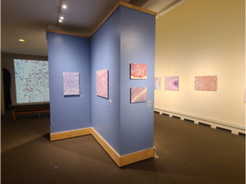
Histological images captured using high-resolution imaging displayed as part of the Death Under Glass exhibition at the Stedman Gallery at Rutgers-Camden.
Beyond its clinical utility, high-resolution imaging serves as a conduit for educational outreach and public engagement. Through projects like the Death Under Glass exhibition, Dr. Hamel and her collaborators leverage captivating imagery to demystify forensic pathology and engage the public in discussions about death, science, and art. By sharing the microscopic world of forensic specimens with a broader audience, Dr. Hamel fosters a deeper appreciation for the beauty of forensic science and its role in society.
Bringing clearer insights to forensic pathology
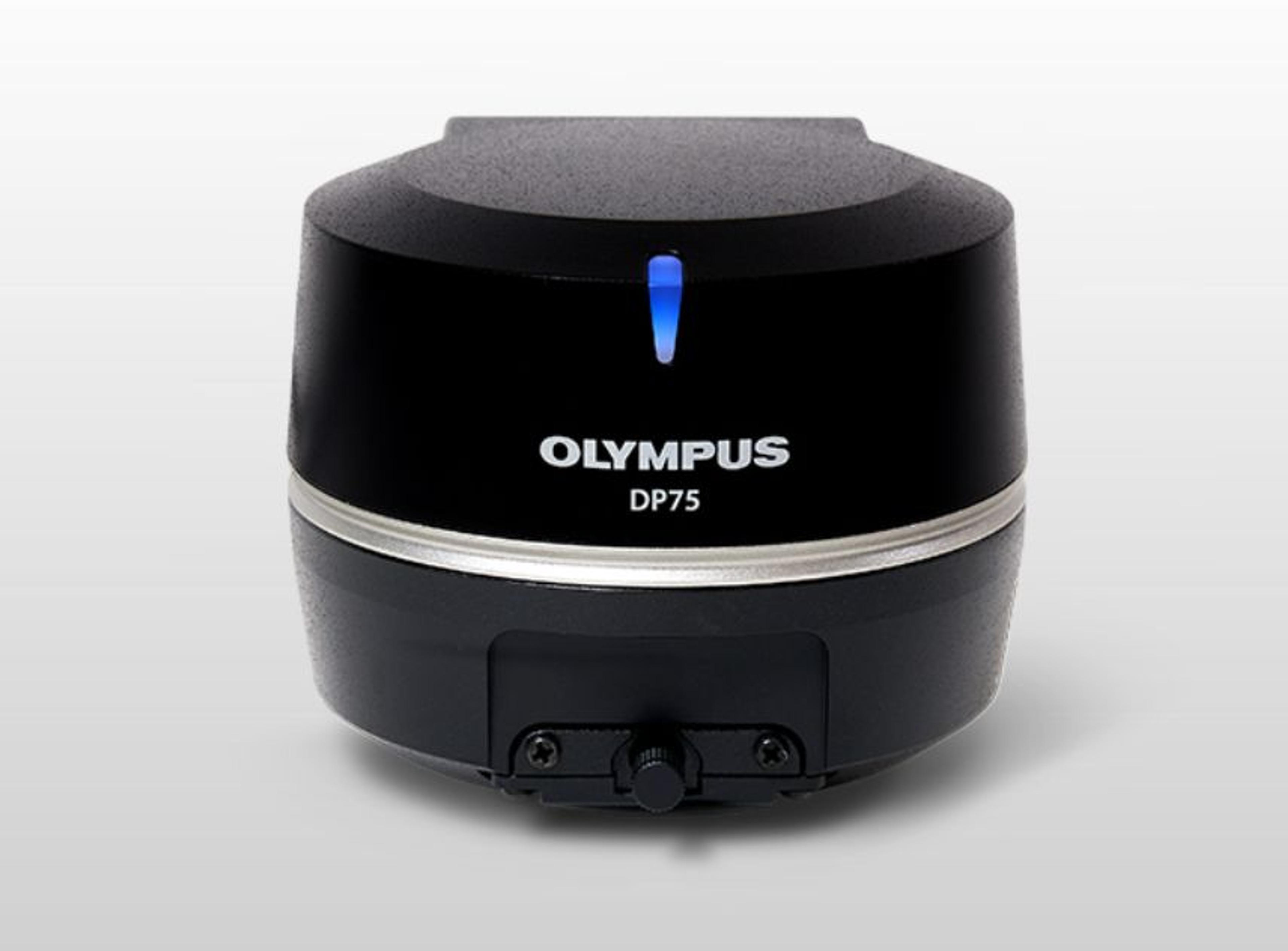
Leading the way in high-resolution imagery in forensic pathology is the DP75 digital microscope camera from Evident.
The DP75 camera boasts a range of features that make it an indispensable asset in forensic pathology laboratories. Its high-performance, multi-application imaging capabilities enable pathologists to capture high-resolution brightfield or fluorescence images with clarity and precision. This versatility enables forensic pathologists to visualize forensic specimens in vivid detail facilitating accurate documentation and analysis.
One of the standout features of the DP75 camera is its exceptional color reproduction, which helps ensure that images are rendered with true-to-life colors, akin to looking through the microscope oculars. This fidelity is paramount in forensic pathology, where accurate representation of tissue morphology and staining patterns is essential for making diagnostic determinations and conveying findings to colleagues and stakeholders. Dr. Hamel remarks, "Thanks to advances by Evident, I'm able to capture what I see through my eyepiece at high resolution and full color, blow it up, and print it on aluminum panels as large as 36 × 24 inches without pixelation or distortion."
Moreover, the DP75 camera supports multiple staining combinations and wavelengths up to 1000 nm with a switchable infrared (IR) cut filter. This capability lets forensic pathologists tailor imaging protocols to specific specimens and staining techniques, helping to ensure optimal image quality and diagnostic accuracy.
The advanced analysis capabilities of the DP75 camera further augment its utility in forensic pathology. Linear mode enables relative intensity evaluation on fluorescence images, facilitating quantitative comparisons of fluorescence expressions. Dr. Hamel emphasizes the camera's ability to capture depth of color and its incredible resolution at high magnification as key factors in its efficacy in forensic investigations. Additionally, the ability to overlay fluorescence and brightfield images with pixel precision enables pathologists to precisely identify the location of fluorescent expression.
The DP75 camera's AI-based scene detection feature is another noteworthy aspect, as it automatically recognizes five observation methods (brightfield, fluorescence, phase contrast, differential interference contrast, and polarization). This intuitive functionality streamlines imaging workflows, enabling even novice users to obtain high-quality images with minimal training.
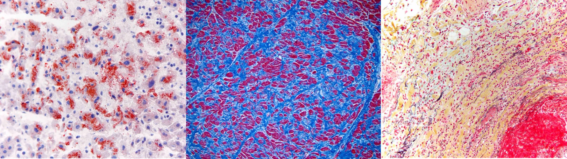
Left to right: Microsteatosis of liver tissue stained with Oil Red O, Masson trichrome stain of the heart, Movat pentachrome stain of blood vessel; all from the @deathunderglass archive
The demand for analytical acumen
Dr. Marianne Hamel, MD, PhD, stands as a luminary in the field of forensic pathology, and her career spans a diverse array of roles and accomplishments. As a per diem medical examiner for both the New Jersey Regional Medical Examiner’s Office in Newark and the Montgomery County Coroner’s Office in Eagleville, Pennsylvania, Dr. Hamel navigates the complexity of death investigations with precision and dedication. Additionally, she is the owner of Jersey Shore Forensics, a private pathology consultation firm specializing in civil and criminal case reviews, private autopsies, exhumations, and cold case work.
The role of a forensic pathologist, as Dr. Hamel points out, is far from mundane, characterized by its challenging nature and the constant demand for analytical acumen. Post-mortem examinations, conducted to determine the cause and manner of death for individuals who die violently, unexpectedly, or outside the care of a physician, require a unique blend of medical expertise, investigative prowess, and unwavering attention to detail. Dr. Hamel's passion for forensic pathology is driven by a desire to solve puzzles and confront the complexities of death head-on.
Through her extensive experience, Dr. Hamel has become intimately acquainted with the indispensable role of imaging technology in forensic pathology. The integration of advanced imaging tools, such as the DP75 digital microscope camera, has revolutionized the way she conducts post-mortem examinations and her research endeavors. In every bit of her work, Dr. Hamel relies on high-resolution imaging to capture the intricacies of forensic specimens, from macroscopic injuries to microscopic tissue samples.
Beyond her clinical practice, Dr. Hamel is a trailblazer in forensic education and public engagement. Her collaboration with artist Nikki Johnson MFA, BFA on the Death Under Glass project exemplifies her commitment to showcasing the beauty and complexity of forensic science through captivating imagery. Through exhibitions and publications, Dr. Hamel shares her insights with the world, bridging the gap between science and art in the process.
Innovations in microscopy equipment
The future of forensic pathology is intertwined with the continued advancement of microscopy equipment, exemplified by innovative technology such as the DP75 digital microscope camera. Dr. Hamel notes, "Innovations in microscopy equipment, such as the DP75 camera, have the potential to revolutionize the field of forensic pathology, empowering pathologists to achieve new levels of precision and insight."
In the coming years, as forensic pathology continues to evolve in response to emerging challenges and opportunities, the role of microscopy equipment will undoubtedly remain central to the discipline. By embracing innovations like the DP75 digital microscope camera, forensic pathologists can unlock new possibilities in forensic investigations, driving progress and innovation in the field.