High-resolution imaging of single PTFE molecules on Teflon surface
Guest editor Vladimir Korolkov, Senior Applications Scientist, Park Systems, outlines a straightforward practical approach for high-resolution imaging
8 Jun 2021
In this guest editorial by Vladimir Korolkov, Senior Applications Scientist, Park Systems, learn about AFM as an important tool in the investigation of the molecular structure of polymers.
Structural studies of polymers are vital since the application of polymer materials ranges from micellar drug carriers to bulletproof vests. Their exquisite molecular architecture provides polymers with a variety of unique properties. The ability to image this molecular structure in real-space is, therefore, critical. The only technique that can perform such imaging is Atomic Force Microscopy. (AFM).
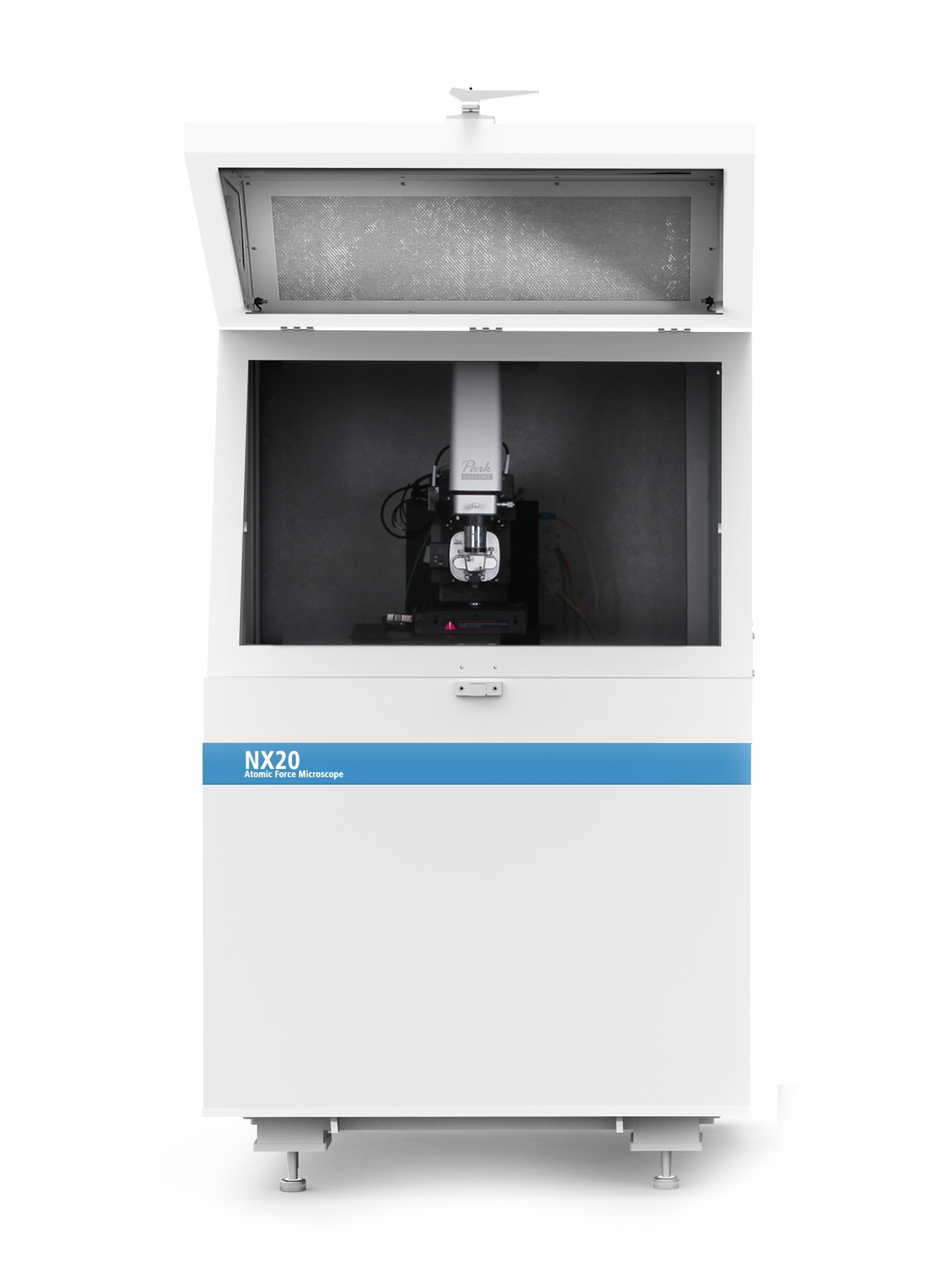
AFM was developed more than three decades1. However, it took a long time to utilize AFM to obtain high-resolution images of polymers. We can certainly mention the development of torsional tapping to observe single polyethylene molecules by Hobbs and Mullin2,3, bimodal tapping by Proksch4 and higher eigenmode imaging by Korolkov5. The latter technique, unlike others, does not require any special cantilevers or custom-modified AFM components. In this work, we have implemented this technique on a commercial AFM – Park Systems NX20 – to achieve molecular resolution on a real-world sample of Teflon.
Polytetrafluoroethylene (PTFE), commonly known as Teflon, is a fluorocarbon solid that has one of the lowest coefficients of friction. Therefore, it is widely used as a material with low adhesion or as an inert coating. Despite its chemical simplicity, PTFE exists in four different crystalline phases that have been studied with electron diffraction techniques6,7. Interestingly, no high-resolution AFM data of Teflon have been published to date.
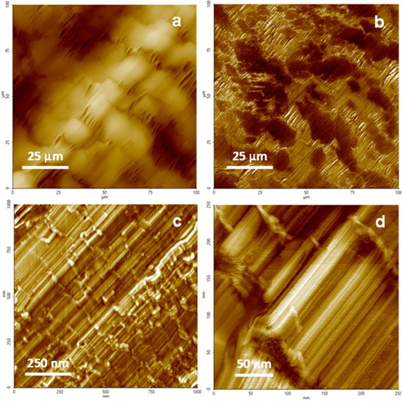
Teflon is known to be a semi-crystalline polymer7. Figure 1 shows a set of large-scale images of a Teflon surface. A 100 µm x 100 µm image (Fig. 1 a and b) already shows two distinctive areas: large 20 µm domains and rope-like areas connecting them. A closer look at domain areas reveals their highly directional nature. A high-resolution phase image (Fig. 1d) shows predominantly crystalline regions separated by smaller amorphous regions on the surface of the polymer. These crystalline domains exhibit flat terraces as observed in Figures 1c and d.
On the following height and phase images (Figure 2), we examine these flat terraces a bit closer. A 100 nm x 100 nm height image (Fig. 2a) shows ~5Å steps with sharp edges. Corresponding phase images (Fig. 2b and c) reveal the true molecular nature of these flat steps – we can clearly resolve single molecules with a period of 5.6Å. A cross-section (Fig. 2d) gives a clear periodic structure of the terrace. From this cross-section, we can also measure the width of a single line at full width at half maximum being 3.5Å. This determines the maximum resolution achieved on this sample. When comparing the observed period of 5.6Å to the reported diffraction data of PTFE unit cell a = 5.66Å7, we can note a remarkable agreement.

Conclusion
To conclude, we have demonstrated a straightforward practical approach for high-resolution imaging using higher eigenmodes of a standard cantilever in tapping mode on a commercial large-scale NX20 Park Systems AFM achieving molecular resolution of a Teflon sample. AFM, a surface-sensitive technique that is always confined to the utmost/outermost surface layer, is able to accurately reproduce results obtained from volume average techniques. Thus, proving that AFM is an important tool in the investigation of the molecular structure of polymers. In fact, AFM is able to provide more structural information by shining a light onto the amorphous regions of Teflon – something that diffraction techniques cannot easily achieve. For instance, a phase image (Fig. 2b) shows that PTFE molecules extend from well-ordered crystalline regions further into the amorphous region of the polymer. The highly localized and high-resolution nature of AFM places it in a unique position to investigate the structure of polymers in real space.
Do you use Park Systems products in your lab? Write a review today for your chance to win a $400 Amazon Gift Card>>
References
Binnig, G., Quate, C. F. & Gerber, C. Atomic Force Microscope. Phys. Rev. Lett. 56, 1986
Mullin, N. & Hobbs, J. Direct Imaging of Polyethylene Films at Single-Chain Resolution with Torsional Tapping Atomic Force Microscopy. Physical Review Letters vol. 107. 2011
Mullin, N. et al. “Torsional tapping” atomic force microscopy using T-shaped cantilevers. Appl. Phys. Lett. 2009
Kocun, M., Labuda, A., Meinhold, W., Revenko, I. & Proksch, R. Fast, High Resolution, and Wide Modulus Range Nanomechanical Mapping with Bimodal Tapping Mode. ACS Nano 11, 2017
Korolkov, V. et. al. Ultra-high resolution imaging of thin films and single strands of polythiophene using atomic force microscopy. Nat. Commun. 2019
Clark, E. S. The Crystal Structure of Polytetrafluoroethylene, Forms I and IV. J. Macromol. Sci. 2006
Brown, E. N., Clausen, B. & Brown, D. W. In situ measurement of crystalline lattice strains in phase IV polytetrafluoroethylene. J. Neutron Res. 2007