Highest Ever Resoultion Images Obtained by Cornell University Researchers Using EM Ptychography
25 Jul 2018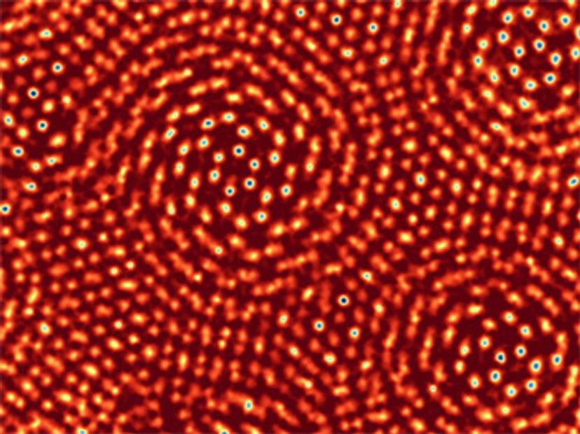
Researchers at Cornell University have used ptychography to achieve the highest resolution ever produced in an electron microscope. The team, led by David Muller, Professor of Applied and Engineering Physics, reported their findings in the July 2018 issue of Nature, outlining their ability to resolve images to a resolution of 0.39 Å. This level of resolution has enabled them to visualise individual atoms with greater contrast, and less damage than was previously possible. To put their achievement into perspective, it represents a 2.5x improvement over the conventional imaging performance of their microscope.
Ptychography is a computational imaging technique that utilises sophisticated algorithms to reconstruct an image from overlapping diffraction patterns, created when an object is illuminated. In their paper, Muller’s group use a powerful new electron camera, which they have developed, to record the diffraction patterns with exceptional sensitivity, speed and dynamic range. The world-record resolution was made possible by the combination of detector and ptychography, heralding a new era in electron microscopy.
Phasefocus™ has commercialised ptychography as its proprietary platform technology, creating a portfolio of products to accommodate a wide range of imaging applications, encompassing life science, healthcare, engineering, metrology and more.
The Phasefocus πbox reconstruction engine, suitable for electron microscopy, is a back-end processing platform that can be connected to a microscope, network or cloud based, to deliver reconstructed ptychographic images to the user’s own software. Compatible with electrons and any electromagnetic wave, πbox deciphers raw diffraction patterns to streamline image generation, making it applicable across a wide range of imaging modalities including X-ray, Electron microscopy and optical microscopy.
Do you use any Phasefocus products in your lab? Write a review today for your chance to win a $400 Amazon voucher!