How Circulating Tumor Cells Are Being Used in Cancer Research
15 Oct 2014
Although often in low abundance, circulating tumor cells (CTCs) have emerged as a potential source of clinically-useful information that can be accessed with far greater frequency than tissue biopsies. In addition, CTCs have a key advantage of being enriched above the background cell levels, enabling them to be analyzed using molecular based methods. An article in Science Daily this month provides a great overview of how circulating tumor cells have provided a genomic snapshot of breast cancer.
The application of next-generation sequencing (NGS) for rare diseases helps to identify new genetic events and understand their molecular pathogenesis through the analysis of CTCs. The IsoFlux System and assay kits, from Fluxion Bioscience, are designed to work specifically with CTC material, significantly increasing the overall target purity of the sample and providing a robust nucleic acid extraction method that is optimized for NGS. The IsoFlux NGS assay kits incorporate a proprietary method of purity enhancement that, when combined with the IsoFlux System for primary enrichment, delivers a high-purity DNA or RNA sample immediately ready for NGS. In this study, CTCs are analyzed using Fluorescence in situ hybridization (FISH) on the IonFlux HT. The SMARTer Stranded RNA-Seq Kit, from Takara, provides a solution for generating libraries for Illumina® sequencing platforms. This kit generates robust data with subnanogram input, as described in this study, making it ideal for the identification of genetic variants in from CTCs in rare cancers.
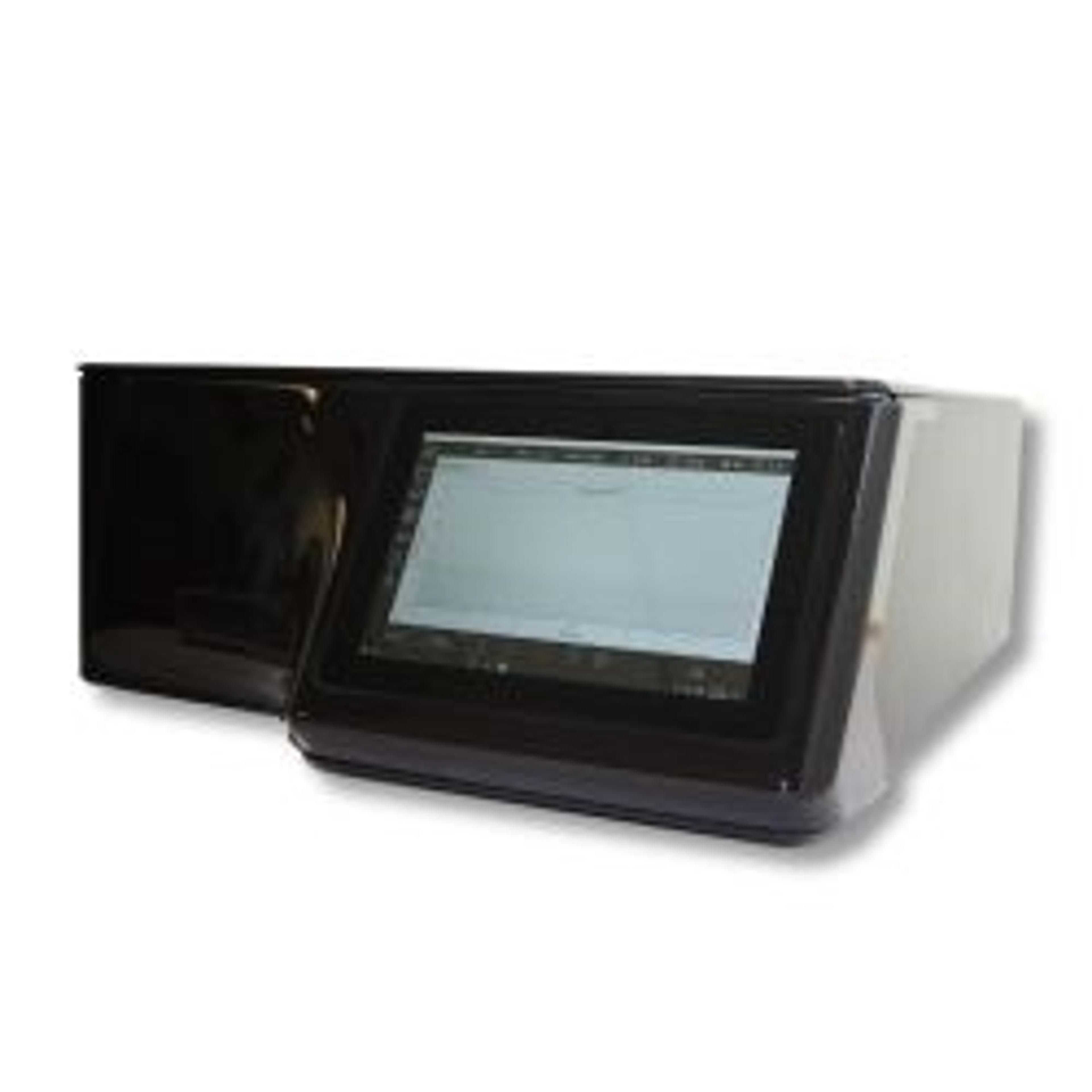
The QIAxpert® instrument, from QIAGEN, can accelerate quantification and quality control of DNA and RNA samples from CTCs for NGS, while the new CLC Cancer Research Workbench is the first complete bioinformatics software suite for rapid analysis, user-friendly visualization, and accurate interpretation of advanced NGS data in cancer research.
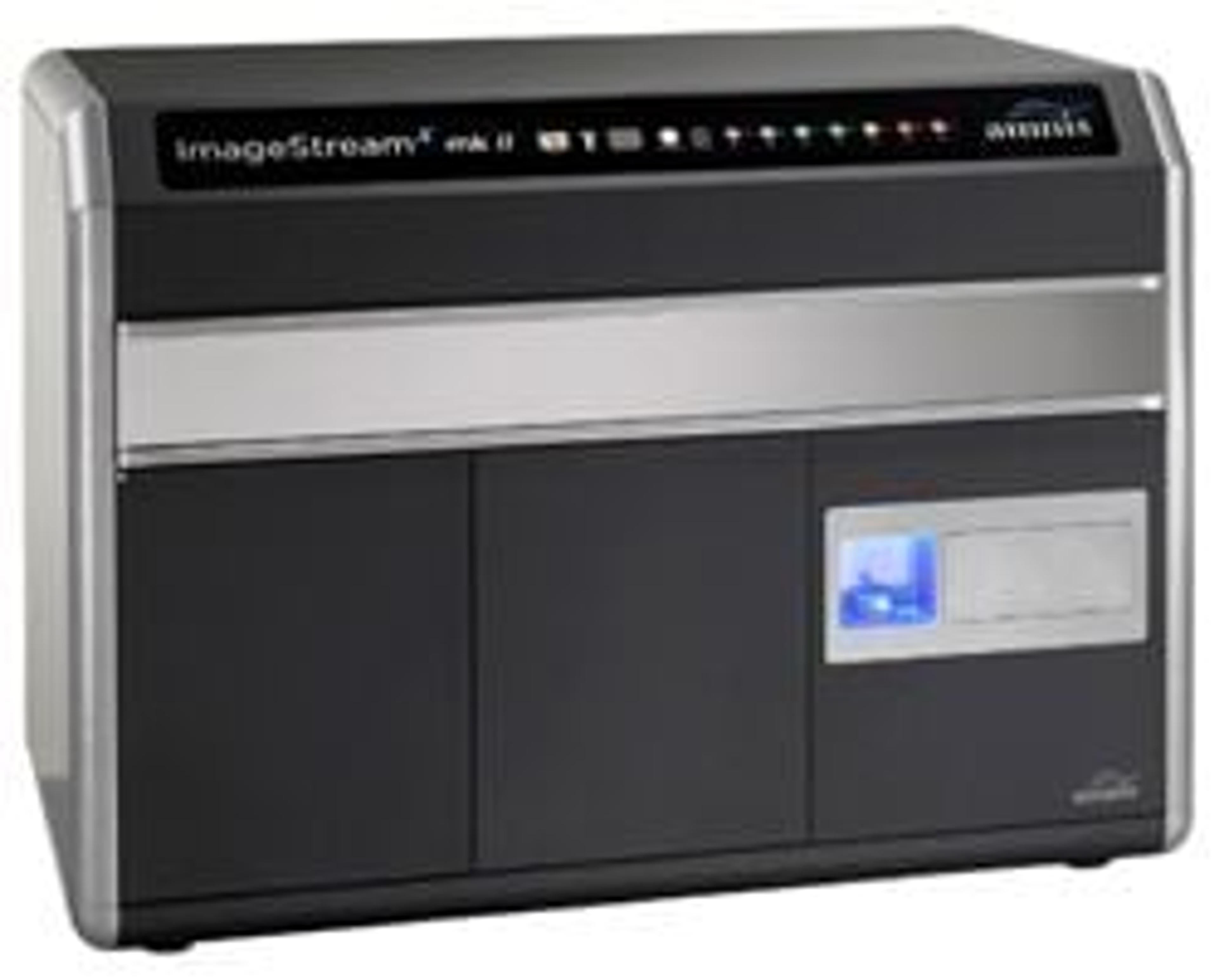
The cell-cell interactions of CTCs and other metastatic cells can be studied using the Image Stream®X Mark II Imaging Flow Cytometer. The high-resolution and sensitivity, combined with up to 10 fluorescent markers, enables the analysis of rare sub-populations of potentially metastatic cells, including CTCs. The instrument combines the speed, sensitivity, and phenotyping abilities of flow cytometry with the detailed imagery and functional insights of microscopy.
The further characterization of CTCs at the molecular level will facilitate personalized medicine via analysis of pharmacodynamic and diagnostic biomarkers. Counting the number of CTCs can also tell us whether a patient's cancer is aggressive, or whether it is stable and responding to specific therapies.