How Mass Spectrometry Evolved into an Indispensable Biological Technique
Dr. Jim Langridge, Director of Scientific Operations for Waters Corporation, shares his scientific journey alongside mass spec technology and reveals what’s in store for the future
1 Feb 2018
Mass spectrometry had its beginnings inside analytical labs, offering reliable profiling of molecules. Not too long ago, it began to occupy a place of value in biological labs as well, as researchers began parsing tissues for molecular signatures of diseases.
In this exclusive SelectScience® interview, we speak with Dr. Jim Langridge, Director of Scientific Operations for Waters Corporation, who has seen and experienced the different phases of mass spec. Read on to learn how mass spec has evolved over three decades, what powerful tools are being developed for scientists and what the future of mass spec looks like.


Dr. Jim Langridge Dr. Jim Langridge, Director of Scientific Operations for Waters Corporation, Manchester UK, is an expert in the field of mass spectrometry, authoring over 60 peer-reviewed publications and 8 granted patents.
SS: As the director of health sciences at Waters, what does your role entail?
JL: As the director, I have the global responsibility for scientific development of technologies and applications around life sciences, and particularly using mass spectrometry (MS) technology. We’re increasingly looking at translating research into assays, such as an LC/MS test, and to verify and validate the performance of these assays in a higher throughput environment to see if they have promise to move towards a more routine end-point.
SS: When did you start using the mass spectrometer? How has the mass spectrometry field evolved since you started working as a scientist?
JL: I got my PhD in biochemistry, and during my doctoral studies I started using mass spec to answer biological questions. I was studying enzyme dynamics then, and during that phase – now about 20-30 years ago – it became clear to me that mass spec was going to make a big impact on biological sciences. The real change that I’ve seen in two decades is the improvement of sensitivity in analytical systems and speed. Traditional biological techniques looked at isolating one compound, one protein or metabolite, using mass spec, to understand the structure and the role of the molecule. But now, the way experiments are done, we can look at a much wider compound class. Rather than simple mixtures, we can now analyze more complicated biological mixtures – proteins, metabolites, lipids – and understand their potential role with regard to a specific disease. So, the field has moved from more of an isolated view to a more integrated view of biology.
SS: What has been your observation of some recent developments in the field of mass spec?
JL: Mass spec imaging is an area that has a great potential – there is a big interest in understanding spatial localization of compounds using mass spec. In this case, rather than taking a whole cellular population and homogenizing it, one would maintain the tissue as in pathology or microscopy approaches, take a slice of the tissue and analyze it by mass spec. By effectively probing each position in an array, you can get a molecular picture of the tissue section analyzed. Such an approach can give us an idea about not only what the molecules are, but also where they’re localized and how they are translocating through membranes, for example. That's something we've put a lot of effort into developing. It’s traditionally been done using matrix-assisted laser desorption/ionization (MALDI) – which we have a product for – but we’re also working with Prosolia in developing desorption electrospray ionization (DESI) for tissue imaging.
SS: What is SONAR at Waters, and how will it help researchers?
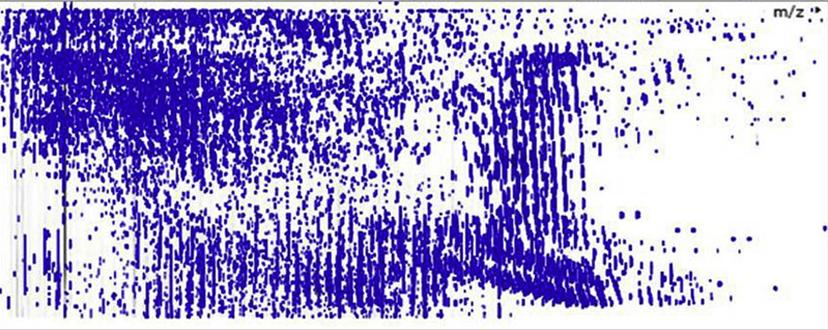
JL: It is a data-independent analysis system. So, rather than the instrument making decisions about which ions to fragment and which species to perform a tandem MS on, the instrument is used in a predictable but unbiased way to acquire data. For example, we characterized the SONAR approach in a recent publication for lipids analysis where a scanning quadrupole is used in front of an orthogonal time-of-flight (TOF).
The new mode of acquisition, SONAR, enables us to get better selectivity, or better sensitivity at the same selectivity. In complex mixtures, one of the challenges is ‘how many components are in there, and am I really sure of what I’m looking at?’ This approach enables us to dig deeper into that sample mix at much faster acquisition rates than previously possible, to allow higher-throughput in addition to selectivity. It will be interesting to see how we progress this in the future, as we are just at the beginning of using this acquisition technique. We’re intrigued as to how the data gets used by researchers to enable better biomarker discovery.
There's been a lot of focus on potential omics research – from discovery of novel markers to understanding of disease mechanisms. We are developing a wide range of different technologies to aid that process.
Dr. Jim Langridge Director of Scientific Operations, Waters Corporation
SS: How does Waters mass spec technology benefit the ‘omics’ field, for example, proteomics, metabolomics and lipidomics?
JL: There's been a lot of focus on potential omics research – from discovery of novel markers to understanding of disease mechanisms. We are developing a wide range of different technologies to aid that process. From a mass spectrometry perspective, we can use that sonar acquisition technique for all three of the application areas mentioned – we can either use it for rapid profiling of complex protein mixtures or metabolomics and lipidomics.
An interesting piece that we’ve done is with the National Phenome Centre, UK, where the goal is to use MS technology to perform large-scale metabolic phenotyping. We’re trying to do two distinct things: one is to look at what the clinical phenotype is, so as to really understand and link clinical conditions to changes at the metabolite level, and the second is to look at epidemiology. Our research question could be: ‘Why do people have a higher propensity for increased blood pressure or increased risk of coronary heart disease?’ We find increasing associations between human metabolites and the microbiome in those specific diseases, and the propensity of an individual for these diseases. From a technology perspective, that comes down to not only selectivity and sensitivity but also robustness and reproducibility. Some of these studies might be 20,000 samples, so you need really good stability of the mass spec platform to make quantitative measurements over long periods of time.
SS: Mass spec has typically been an analytical technique that has recently seen growth in translational research. What are your views on how mass spec can help with biomarker discovery?
JL: The promise of biomarker research is to identify subtle changes in key proteins, a panel of proteins, metabolites or lipids, so as to allow identification of early-stage disease states, and for better therapies to be targeted towards certain populations of patients. Today, we treat all patients with the same type of drug. If they have, for example, a different type of cancer, traditionally it would be one type of treatment. But now, using mass spec technology and characterization, we can further stratify patients even past the four accepted molecular subtypes for colorectal cancer. This can enable precision medicine where different treatment regimens for each of the stratified patient populations can be applied. We are just scratching the surface of such discoveries. As we understand more about molecules in diseases and the way to differentiate them — not just based upon a genetic profile, but also based on proteomics and metabolomics profile — more specialized, personalized treatments will be developed.
SS: Can you share an example of such a project?
JL: One project I would highlight is the collaborative work with Prof. Louise Kenny at the Infant Centre in Cork, Ireland, that focuses on preterm birth, a serious issue that is on the increase. We’re trying to build up a profile of metabolic markers that would predict whether a mother was going to have a spontaneous preterm birth or not. If a panel of biomarkers is developed, potentially, they could treat an at-risk mother and that can save both the mother’s and the baby’s life. The project is in the early stages, but if the research studies are carefully controlled then, at some point, they can be translated into an assay to stratify at-risk patients.
Useful resource for mass spec scientists
Have you checked out our 'How to Buy Mass Spectrometers' eBook? Get instant access to the eBook here.
SS: What is the future of mass spec technology?
JL: The future lies in hyphenating multiple technologies. LC and GC were two standalone separation techniques that later came to become hyphenated with mass spec as LC/MS or GC/MS. Now, we’ve hyphenated MS with ion mobility as well, i.e. separating ions in the gas phase by their shape or size or charge state.
MS is a very powerful tool, but it is only as good as the sample you put in. So, sample preparation is critical, and the ability to separate quickly using orthogonal techniques will continue to be important in the future.
In addition to fundamental improvements in performance, such as accuracy and selectivity, a very significant next step for the field is rapid evaporative ionization mass spectrometry (REIMS) or what is commonly known as the iKnife, a handheld device that analyzes samples and provides a molecular profile of the sample within seconds. Taking the plume from an electrosurgical diathermy knife and sampling the vapor produced by the tissue directly into the mass spectrometer will provide researchers and medical professionals with a quick readout in the future.
Find out more about Waters’ systems here.
Do you use mass spec in your research? We want to hear from you! Share your expertise here.


