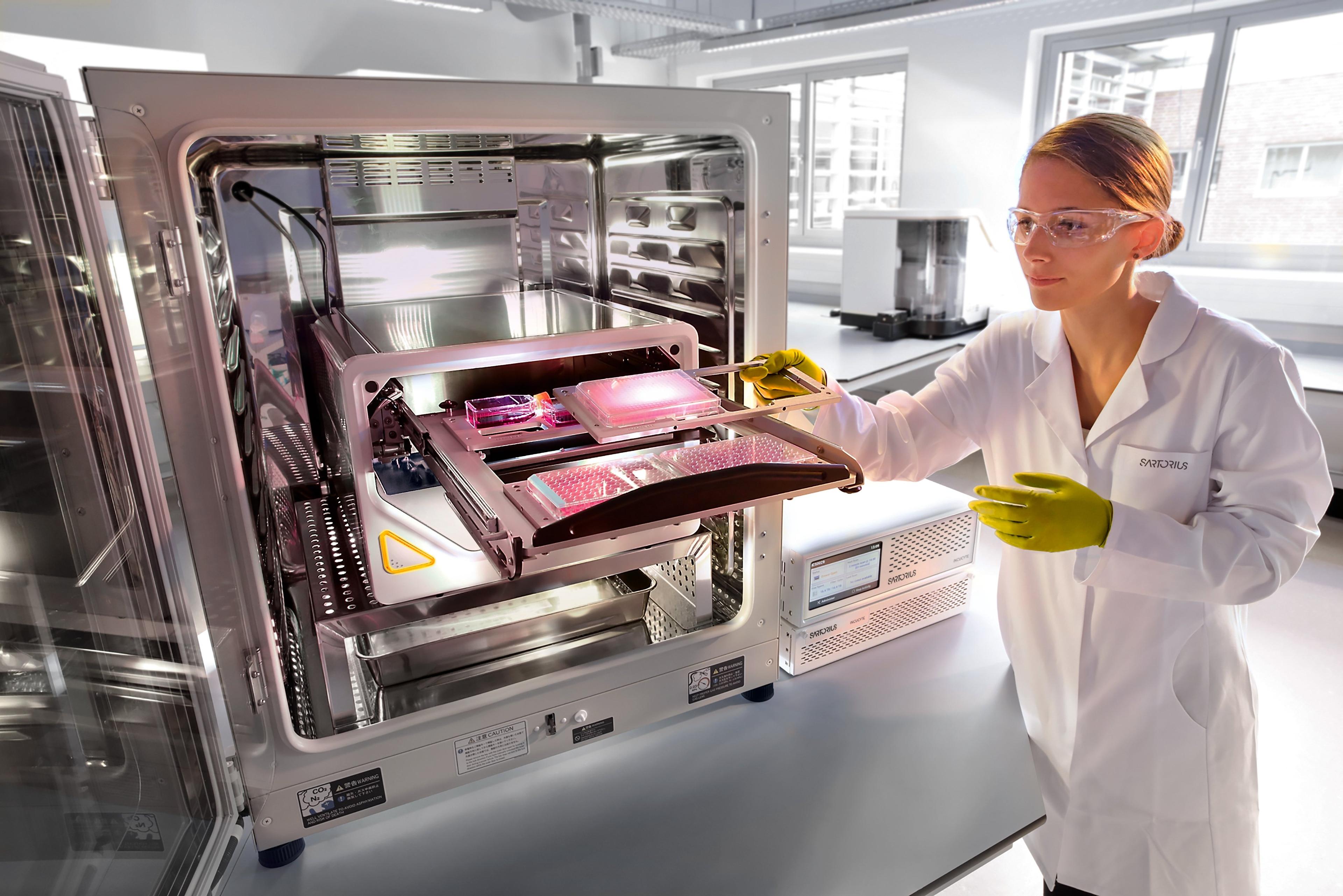
How to ensure confidence in your advanced cellular models with live-cell monitoring
Find out how live-cell monitoring can provide key quality control information during assay set up and discover the importance of cell model optimization for drug treatments
21 Jun 2020

Recent innovations in cell source, manipulation, and assay readout have enabled researchers to develop advanced cell models that better recapitulate the in vivo cell environment within the lab setting.
These complex models pose new challenges in cell analysis, and while cell-based assays have also become technically more complex, the need to holistically capture dynamic, and sometimes subtle, cellular events has become increasingly important to optimize conditions.
This on-demand SelectScience® webinar explores how, by combining real-time imaging data of cellular events with automated quantification, and without disturbing the sample during the entire cell culture workflow, live-cell imaging can support a better understanding of the biological processes at play and direct the optimization of these advanced cellular models to ensure confidence in results.
Think you could benefit from this webinar, but missed it? You can now watch it on demand at a time that suits you and find highlights from the live Q&A session below>>
Watch on demandQ: How long does it take to image the entire 96-well?
This depends on the number of images per well and channels collected. For reference, one image/well and one channel would be around 5 mins per plate.
Q: During live-cell culture and monitoring, does the staining of live-cells with specific antibody marker affect the cellular viability and function? How long can the live cells with antibody live for further experiments?
The effect of an antibody is very antibody dependent. Some will cause cellular effects, but others do not. We have used labelled antibodies in a weeklong immune cell killing assay with no issues and then have used these cells in follow- on flow experiments.
Q: Can you perform quantification on images collected in the flask prior to cell seeding as part of the workflow?
Yes, this can easily be incorporated into the workflow.
Q: Do you need an alternative analysis module for 3D?
Yes, this would be the Incucyte spheroid analysis module.
Q: Can you track distinct populations in 3D co-culture models?
Not using bright field, however with the multi-spheroid application you can potentially track flu labelling cell types in co-culture.
Register to watch the full webinar>>
SelectScience runs 3-4 webinars a month across various scientific topics, discover more of our upcoming webinars>>

