How to Use Live-Cell Analysis for Neuroscience Applications
Learn how live-cell analysis techniques can benefit your neuroscience research, in this expert on-demand masterclass
29 Mar 2019
Despite exciting recent developments in neuroscience, identifying novel, truly effective treatments for patients with neurological and psychiatric conditions remains highly challenging. There is an unmet need for more translational models and advanced analytical techniques that afford greater biological insight and productivity.
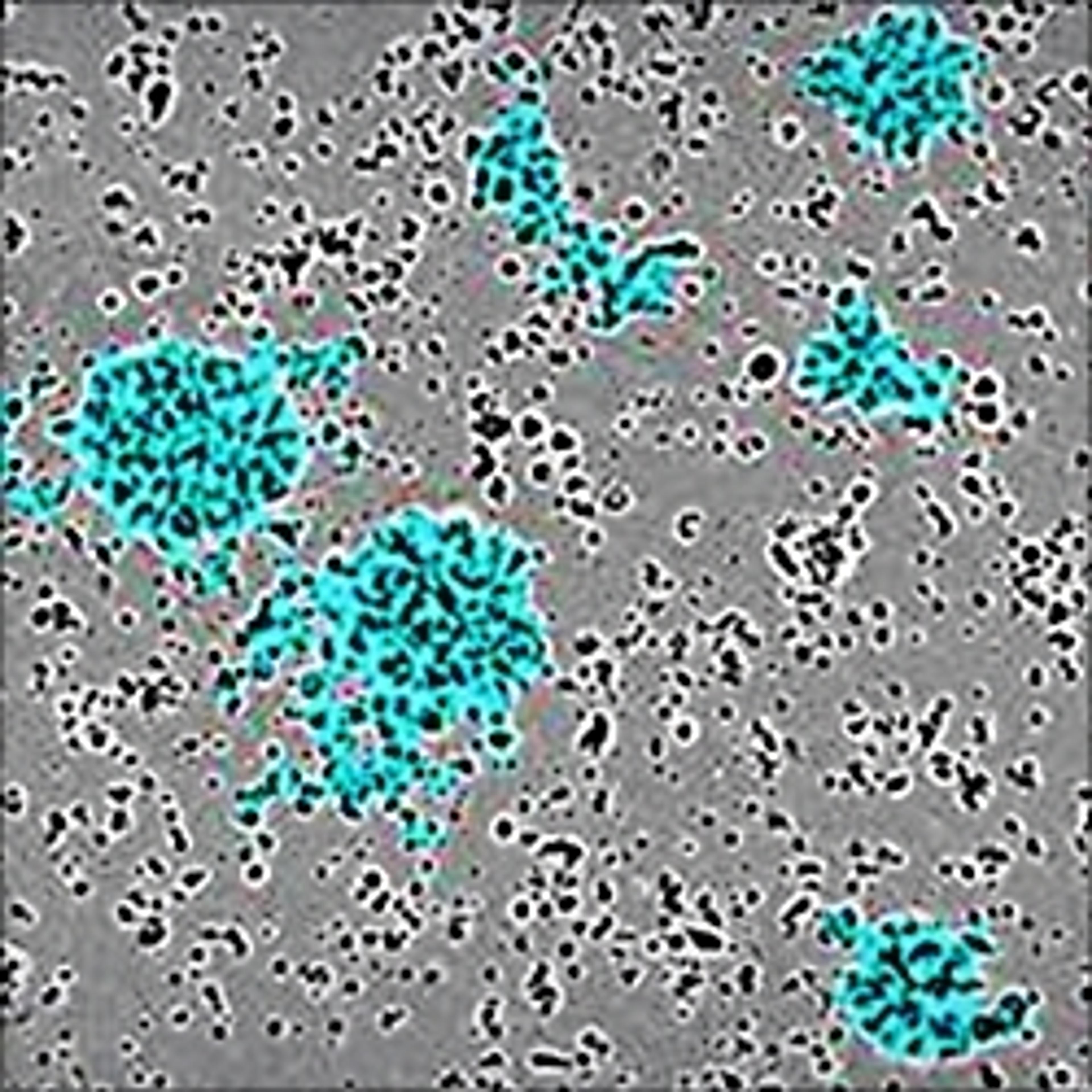
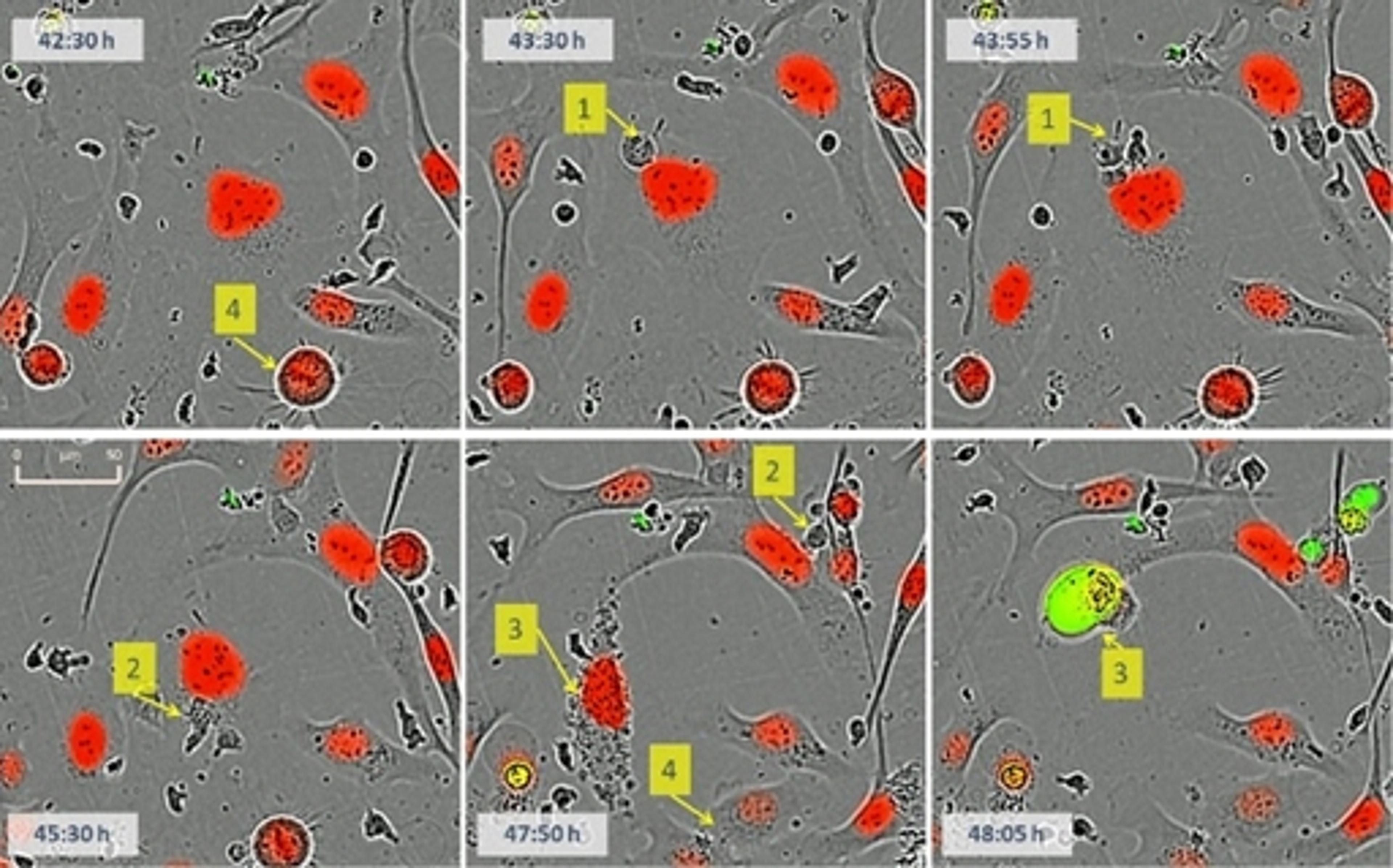
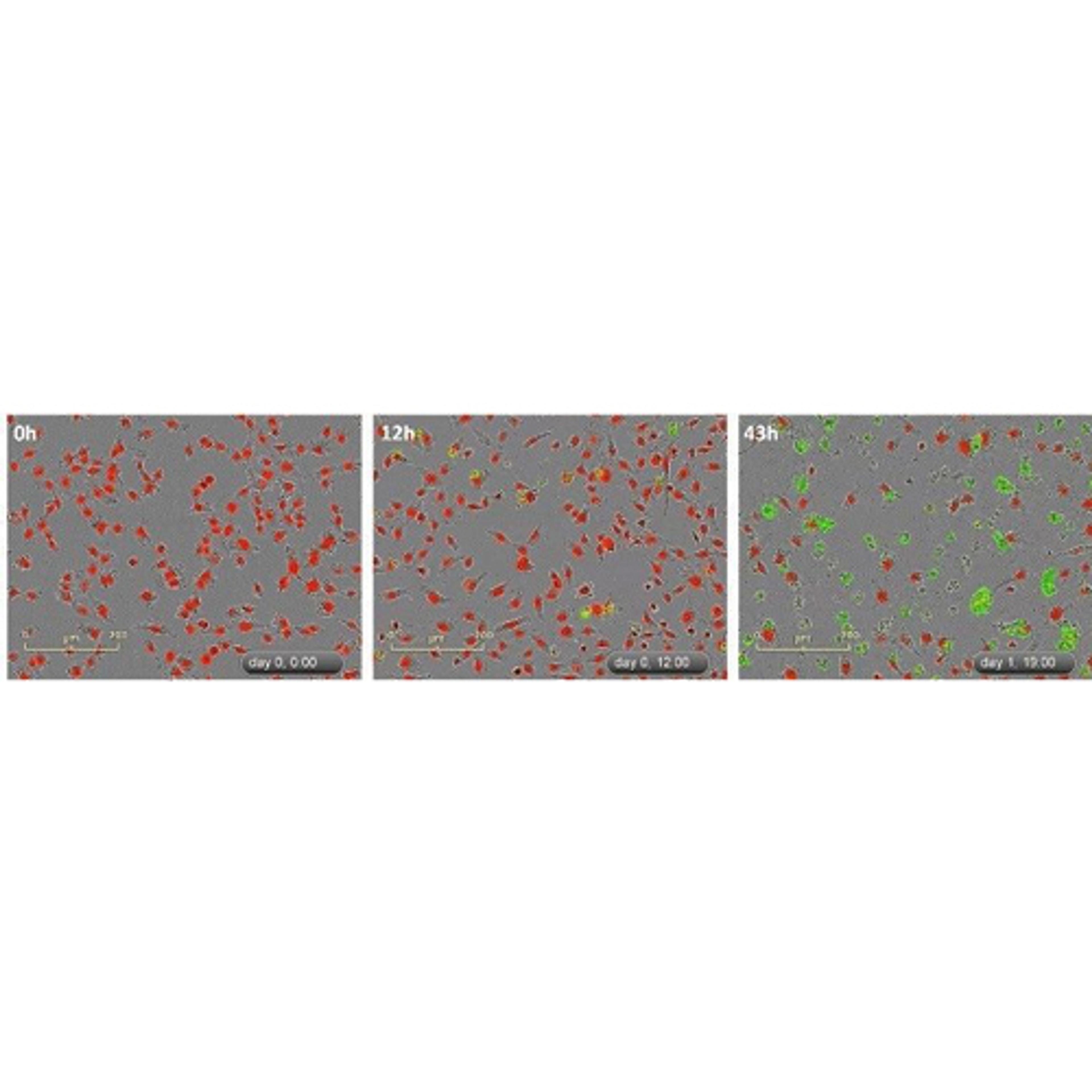
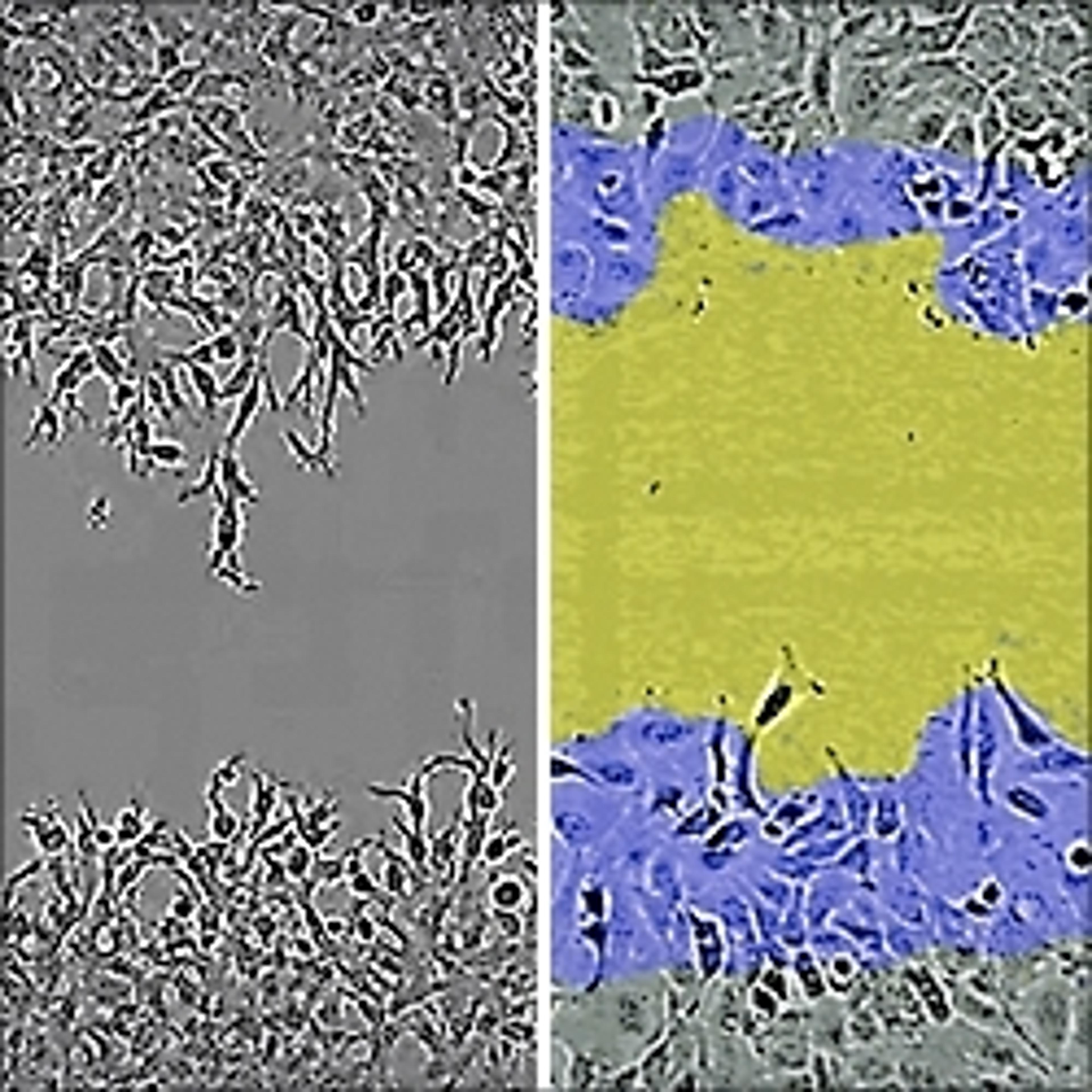
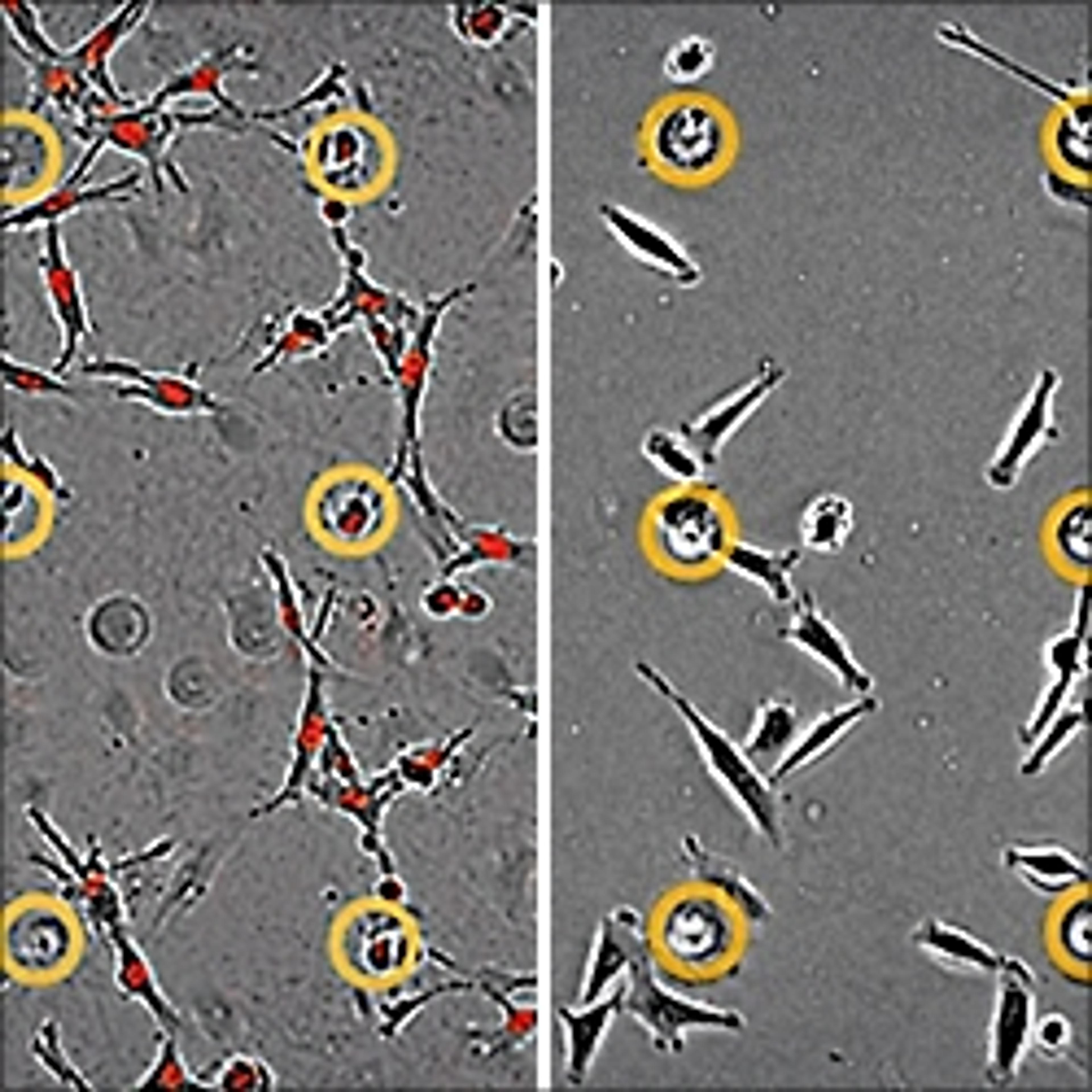
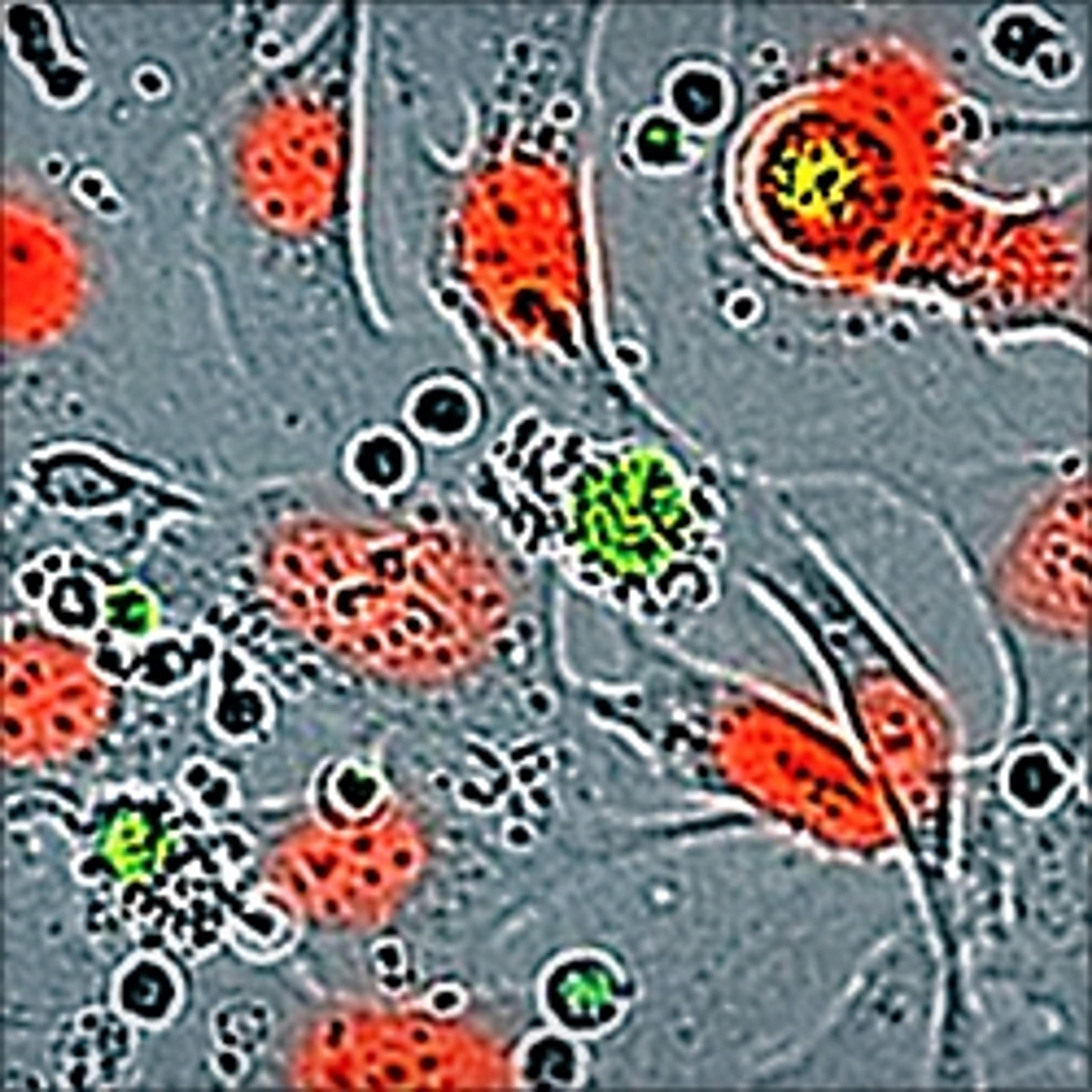
In a recent webinar, now available on demand, senior R&D scientist Susana L. Alcantara discusses novel live-cell analysis techniques and their applications in neuroscience. Watch this webinar to learn how to gain greater biological insight and productivity with advanced cell models, explore examples for neurotoxicity, neurodegeneration and neuroinflammation, and discover how automated assays are used for measuring neuronal activity, neurite dynamics, cell health and neuro-immune functions (phagocytosis, chemotaxis) in stem cells and primary neuronal cultures.
If you missed the webinar, you can now watch it on demand at a time that suits you. Read on for highlights from the webinar’s Q&A session that took place live on Twitter.
Q: iPSC-derived microglia offer huge potential. Have you observed variations in the functional profiles when investigating microglia from different sources?
SA: Yes — the live-cell analysis approach offers insights into the kinetic profiles which do differ between iPSC-derived microglia.
Q: Have you performed end-point ICC to confirm the validity of the neurite length measurements?
SA: Yes, and we have observed a good correlation with ICC using antibodies targeting tubulin. A good example of combining live-cell analysis with an end-point readout.
Q: Would it be possible to image cells growing on a culture inlay in a 24-well plate? The cells would be above the normal plate bottom growing on the inlay.
SA: Currently no, but there is potential. Email askascientist@sartorius.com for more information.
Q: Does the IncuCyte acquire the images during the calcium imaging? Or does it just provide the numbers?
SA: All fluorescent images are captured, stored, and analyzed. Each acquisition is viewable as a movie in addition to the quantification.
Q: For comparison of neurite dynamics, were different neuronal cell types from rodents compared to differentiated human iPSC cells?
SA: Yes, and differential profiles were observed between iPSC from varying sources and rat primary neurons.
Missed the webinar? Watch it on demand now>>
SelectScience runs 3-4 webinars a month across various scientific topics, discover more of our upcoming webinars>>

