Incidence of Free Light Chain Escape in Multiple Myeloma, Why is it Increasing?
Mark Drayson discusses free light chain escape at the 7th International Binding Site Symposium
26 Nov 2015
Mark Drayson discusses the incidence of free light chain escape, a type of relapse seen in Multiple Myeloma patientsMark Drayson Mark Drayson is Director of the Clinical Immunology Service (CIS) at the Medical School University of Birmingham, UK. Mark contributed to the development of Freelite and its clinical validation and has developed a second generation of FLC tests including a rapid point of care test.
In his presentation, delivered at the recently held 7th International Binding Site Symposium, Mark Drayson discussed the incidence of free light chain escape, a type of relapse seen in Multiple Myeloma patients.
Four types of relapse are commonly seen with Multiple Myeloma. At the beginning of his presentation, Mark Drayson described a study in which 35% of the study participants showed an increase in both paraprotein and free light chain levels, 50% showed only an increase in paraprotein levels, 5% showed a small or no increase in paraprotein and free light chains and 10% relapsed with an increase in their free light chains only.
Light Chain Escape
Multiple Myeloma patients who relapse with an increase in free light chains only, without a corresponding increase in intact immunoglobulin levels, are referred to as having light chain escape.
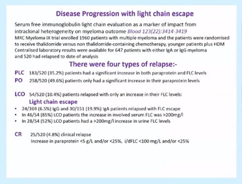
Slide showing the four types of relapse seen in Multiple Myeloma patients
Multiple Myeloma patients with light chain escape have a poorer prognosis than patients who relapse with intact immunoglobulins only. Early laboratory detection of light chain escape means that unnecessary complications, such as renal impairment, can be avoided.
Improved understanding of MM
It was historically believed that Multiple Myeloma was the result of a single abnormal clone; however assay improvements mean that we now know that a whole range of different plasma cell clones are involved. In the example shown in Figure 1, you can see that the clones detected at relapse and disease progression may be different from the clones detected at the point of diagnosis. In the example shown, free light chain only patients showed no change in the type of clone detected, but 44% of the remaining patients changed their paraprotein pattern at relapse.
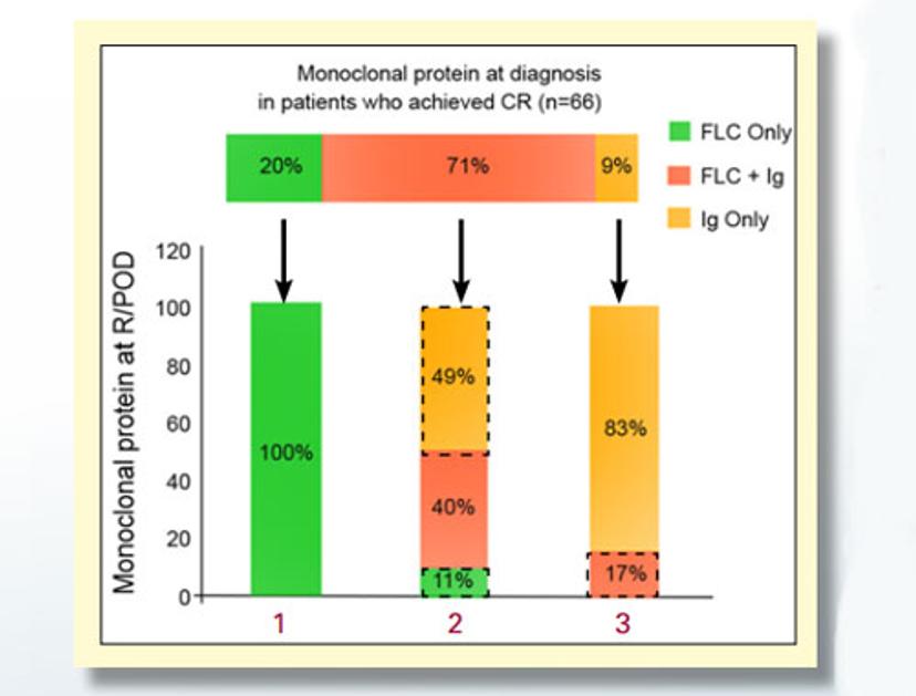
Figure 1: Clones detected at relapse and progression
Laboratory detection of Light Chain Escape
Light chain escape can be detected using Freelite® free light chain assays. During his presentation, Mark Drayson demonstrated that the half-life of free light chains is measured in hours, whereas the half-life of intact immunoglobulins is approximately 20-30 days. This means that free light chain detection is able to give a much faster indication of response to treatment than measurement of intact immunoglobulins.
Developments in the detection of intact immunoglobulins and free light chains have improved our understanding of Multiple Myeloma. These developments have also led to improved sensitivity of detection of abnormal clones and enabled us to detect these clonal changes during patient relapse and disease progression.
It is therefore not necessarily the case that there is an increased incidence of light chain escape in Multiple Myeloma patients, rather that accurate, sensitive and rapid methods of free light chain quantification and characterization have led to increased detection of the light chain escape phenomena.