Intracellular architecture: Creating a map to examine how cells are internally organized in space and time
Learn how researchers at the Chan Zuckerberg Biohub are mapping the human cell to generate an open-source, interactive library
11 Apr 2022
Dr. Manuel Leonetti, CZ Biohub
While we know what proteins make up our cells, how these proteins are expressed in time and space and how they interact with other cellular components aren’t fully understood. Learning more about cellular function and make-up can offer insights into what goes wrong in a diseased state.
Researchers at the Chan Zuckerberg Biohub (CZ Biohub) are setting out to do exactly that: examining the fundamental workings of the human cell. In doing so, they hope to unravel the underlying mechanisms contributing to diseases. The center brings diverse experts from Stanford University, the University of California, Berkeley, and the University of California, San Francisco, along with international collaborators, to collectively find answers to long-standing questions in cell biology.
In this SelectScience® article, we speak with Dr. Manuel Leonetti, Group Leader, Quantitative Cell Science at CZ Biohub, whose research focuses on developing an atlas, called OpenCell, that maps the internal architecture of the human cell.
Mapping the intracellular architecture
The Quantitative Cell Science project is aimed at deciphering how the basic building blocks of the human body are organized and how they communicate with each other. Specifically, the goal is to understand and uncover the cellular physiology in healthy tissue so that it’s possible to better understand, diagnose, and treat diseases caused by cellular disruptions.
“There are two broad themes of research at CZ Biohub’s Quantitative Cell Science initiative: cellular localization and cell dynamics,” shares Leonetti. “Some of us are interested in understanding how tissues are comprised of single-cell units, while some others explore how different cells and tissues come together during development.”
“In my lab, we ask architectural questions at the level of a single human cell,” Leonetti continues. “We’re trying to build an intracellular map of all the proteins that make up the cell.”
Mapping the architecture of a healthy human cell and knowing how it’s built provides the necessary information to predict how it might behave during disease. “Using this cellular atlas, we aim to build predictive models to assess the impact of individual mutations that are very common in patients but aren’t currently linked to a specific disease phenotype,” adds Leonetti. “Having a complete understanding of how the cell is internally wired is a necessary step to studying the cellular perturbations in disease that ultimately manifest as phenotypes.”
Studying protein localization and interactions inside a cell: The technological workflow
Genome sequencing data already provides a list of proteins that make up the cell. However, this list doesn’t answer two crucial questions: “The first is: where is that protein localized within the cell? And the second is: what kind of molecular interactions does a given protein have with other proteins in the cell?” Leonetti notes.
The cell proteome is essentially an interactive, dynamic network of proteins in constant communication with one another. Therein lies the challenge. To map these proteins, the Leonetti lab needs to use technologies that can capture their profiles in time and space.
“To deconstruct the cell one protein at a time, we turn that protein fluorescent using GFP. Today, we can do this well with the surgical precision of CRISPR. So, we're not changing anything in the cell, we're just turning one protein fluorescent at a time,” Leonetti explains. “This allows us to sort and isolate the engineered cells using cell sorting. We can then use live-cell microscopy to examine where the protein is localized within the cell. At the same time, we can also use this fluorescence tag as a target for antibodies. We can lyse the cells to capture the GFP-tagged protein using immunoprecipitation and perform mass spectrometry to determine protein-protein interactions.”
TECHNOLOGY SUITE USED TO CREATE THE CELL ATLAS
- CRISPR gene editing: Fluorescently labeled cell lines
- Automation: Scale experiments and increase throughput
- Flow cytometry: Sort and isolate cells
- Live-cell imaging: Map proteins in time and space
- Mass spectrometry: Profile protein-protein interactions
Using automation to scale research
With over 20,000 different proteins encoded in the genome, coupled with the fact that not all proteins are expressed at the same time, the cell mapping project can quickly become time-consuming and tedious. “This is a challenge that the entire field is experiencing today – being able to scale up our experiments,” notes Leonetti. “We’re using powerful technologies that can answer complex research questions, but the challenge is to apply these technologies at scale, tens of thousands of times, and still get accurate, reliable results.”
A lot of the original intracellular architecture work in the field was done manually. In recent years, researchers are reevaluating how experiments are being performed so as to maximize output with limited time and resources. As such, automation is a sought-after tool in these workflows. “I was very fortunate to be able to work with a team of engineers here at the CZ Biohub to build robots and software to make our lab more productive, and our work less tedious,” shares Leonetti. “My colleague Rafael Gómez-Sjöberg and his bioengineering team are key partners in all of this. An interesting project that we worked on was automating the cell sorter. For that, we also received valuable support from the Sony Biotechnology team.”
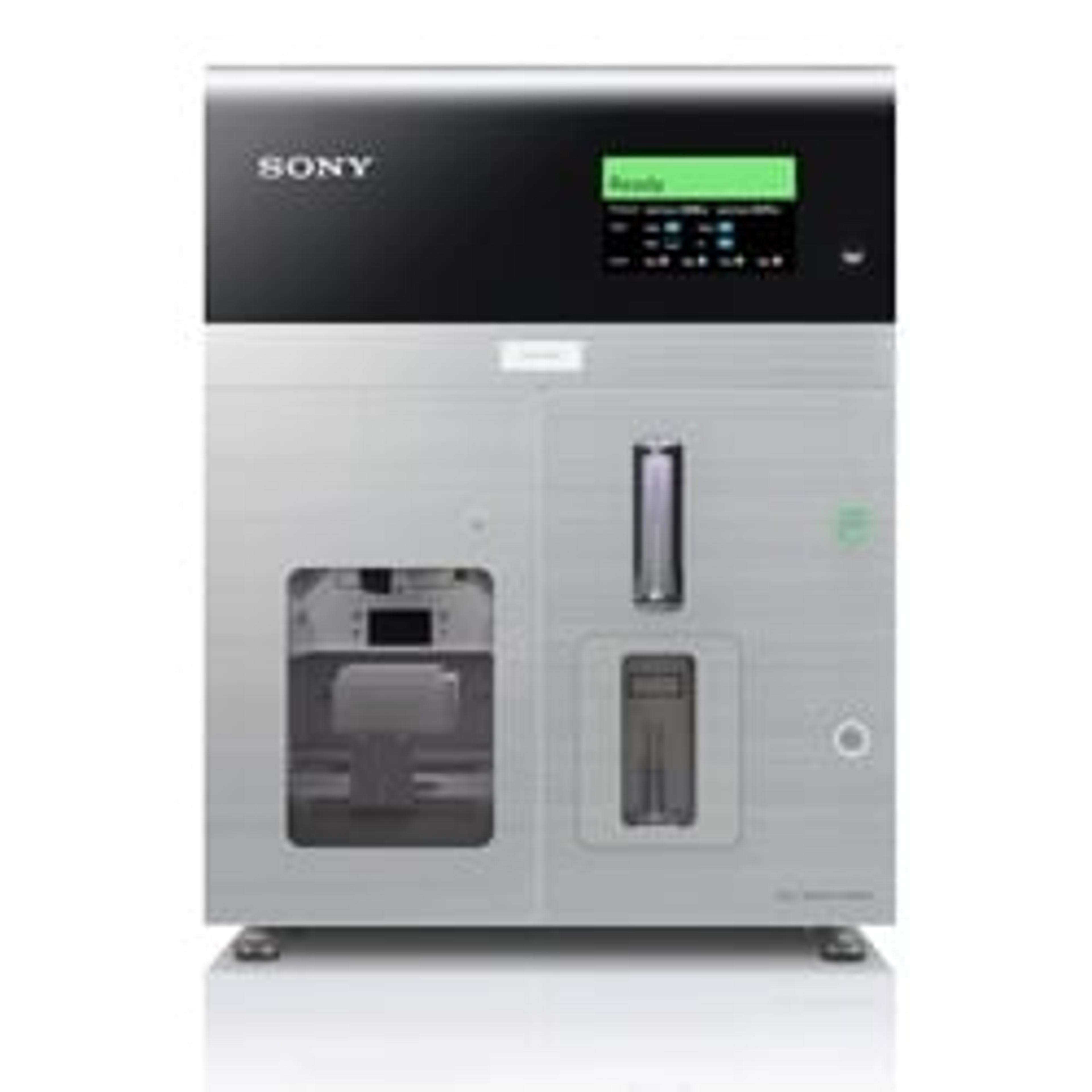
The Leonetti group currently uses the SH800S Cell Sorter to automate the lab’s cell sorting protocol, starting with set-up, acquisition, sorting, and finally, data analysis.
“The field of life sciences is being transformed by innovative technologies, but there is an opportunity cost of not being able to scale technologies,” notes Leonetti. “One of our main objectives is to automate workflows and make them more efficient, cost-effective, and scalable.”
Collaborative efforts to create the open-source data hub: OpenCell
Automation generates a large volume of data which, in turn, poses new challenges – and brings opportunities – for data curation and sharing. “We need to invent new ways of sharing data, making sure that it’s usable and accessible to the entire community,” Leonetti continues. “For us, that means building interactive websites so that everybody can use our data.”
The lab’s flagship project, OpenCell, created from the cellular atlas mapping work, is available for everyone to access and use. It contains an open-access database of ~1300 tagged, localized proteins and ~30,000 protein-protein interactions, providing a systems-level map of the human proteome.
The OpenCell project is a result of the collaborative efforts of different colleagues at the CZ Biohub and across the industry, in particular, a partnership with Prof. Matthias Mann's lab at the Max Planck Institute in Germany is driving innovations in mass spectrometry.
“Collaboration is absolutely essential, especially in a multidisciplinary field such as life sciences. We need different kinds of expertise beyond biology to tackle these complex problems,” says Leonetti. “In addition to collaborating with other research labs, we also work closely with data scientists, software engineers, web developers, and technology providers. To make our work more accessible to the scientific community and go seamlessly from prototype to product, these collaborations are invaluable.”
In the future, the Leonetti group plans to transition from in vitro models to ex vivo models, using cellular systems such as 3D organoids to mimic diseases in a dish.
Discover how novel technologies are driving our understanding of the human cell in this video >>