Is that a live cell or cell debris? Cell viability meets product purity
Watch this on-demand webinar to learn to how to measure cell viability and foreign particle impurities quickly and accurately, in a single workflow
22 Nov 2021

Measuring cell viability is an incredibly important step across all applied cell biology, including cell line development for biotherapeutics and cell immunotherapy. However, traditional tools such as flow cytometry and hemocytometry struggle to differentiate between live cells and cell debris and other contaminants due to their workflows.
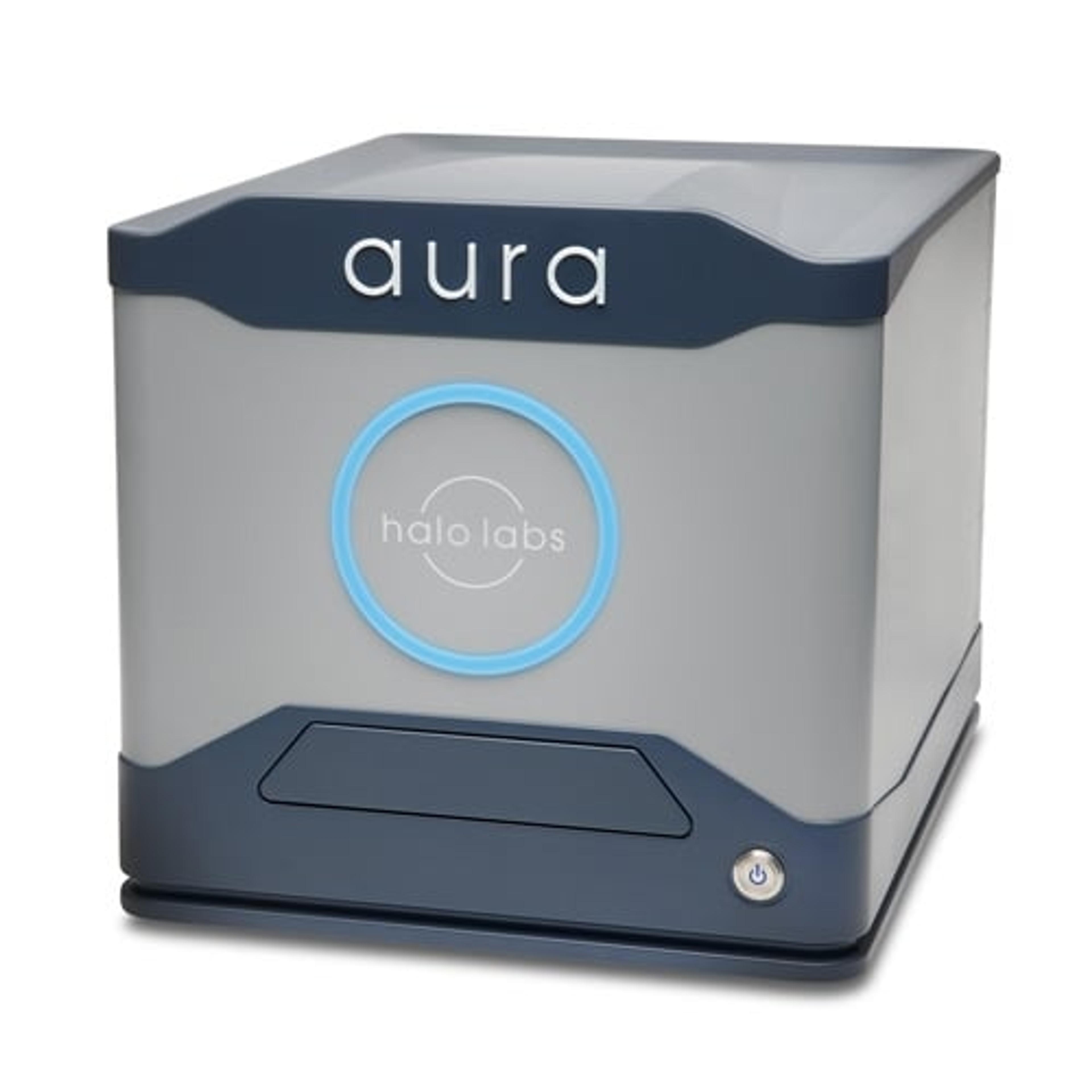
In this free SelectScience® webinar, now available on demand, Dr. Bernardo Cordovez, Chief Science Officer and Co-Founder of Halo Labs, details the benefits of high-throughput, backgrounded membrane imaging and fluorescence membrane microscopy, and shares how these techniques can be used to find residual particulate contaminants such as expansion beads, cellular aggregates, and others in concentrated cell therapy solutions.
Watch on demandRead on for highlights from the Q&A discussion and find out how to increase the accuracy and speed of your cell viability and foreign particle impurity measurements.
How does the sampling efficiency of Aura compare to MFI and FlowCam?
BC: One of the great advantages of this system is that it has 100% sampling efficiency. Typically, capillary-based systems, whether flow cytometry, flow imaging, or light obscuration, miss lots of particles because of the low refractive index contrast.
A second advantage is that, whether you have a focus microscope objective to take images or you focus laser light to interrogate particles, the focal depth of any objective is limited. You have 4x objective-only images in focus in around 40 microns depth of focus, but the channels tend to be much taller than that. So, the true sampling efficiency is in the order of, say, 20% of what is being measured that is in focus.
In the case of the BMI, 100% of the samples were filtered through a known well and 100% of the samples were in focus. So, we have 100% sampling efficiency, with everything is in focus, while the true sampling efficiency of high-quality detection is in the order of 20%.
Do I have to stain on the membrane or in solution?
BC: It depends on the workflow. For a lot of the viability cellular workflows that we discussed, some of the staining is done in solution. There are advantages to that, as there are some kinetics that you can exploit. For more purity/sterility-type questions, it's best to stain on the membrane. You get some really nice surface-based kinetics that have very low incubation times, which seems a little bit counterintuitive as when you stain particles, they filtrate directly on the membrane. The nice thing about that is that, for example, if your dye was, for some reason, a little bit insoluble, you wouldn't measure those particles because you're decoupling your particle detection, your imaging counting and sizing versus the fluorescent stuff if you do the incubation on the membrane. So, you can do both.
They depend on the workflow. Typically, our go-to is to stain on the membrane, but for some of these cellular assays, we do it in solution.
What fluorescent dyes are compatible with the Aura?
BC: We are compatible with a lot of the Alexa dyes, for ADA, so you can do antibody-based labeling; thioflavin T for protein detection; a lot of the blue dyes for your nucleic acid staining, such as DAPI and Hoechst; and there are also lipid dyes like BODIPY. So, it's a whole slew of compatible dyes.
So, you can use any cellular protein or lipid dyes of choice, and it comes with some of the most common fluorescence channels. It's a platform where you can use pretty much any dye to interrogate your chemical or biological question you want to ask.
Can Aura be used to measure lentiviral aggregates?
BC: Yes, this is one of our key applications within cell therapy. Lentiviruses are one of the most important working materials for transfecting T cells or NK cells because they can do large genome modifications that you can't do with AAV. It's also incredibly inexpensive, so it's in high demand. There's lots of either AAV which we measure, or any other virus or protein particle, and it tends to aggregate, so measuring it is very important. One of the fastest-growing places for this technology within cell therapy is to qualify and measure lentiviral aggregates.
For cell therapy, what does it mean to have many large cellular aggregates?
BC: Cellular aggregates can be problematic. Cell therapies are injectables, so have to meet USP standards. If cells are on the order of, 8-10 microns per T cell, if you have very large clusters of cells, these can present a risk for capillary occlusion, and a high immunogenicity. So, a perfectly efficacious cell therapy might be rejected by the body.
With gene therapies and cell therapies, their formulation development is in its infancy. Only a handful are release products. This tends to present a lot of therapies with highly aggregated particles, and that needs to be measured because big particles are dangerous.
To learn more about measuring cell viability watch the webinar on demand >>
SelectScience runs 10+ webinars a month across various scientific topics, discover more of our upcoming webinars>>

