Leveraging ZEISS Airyscan to Integrate Subcellular and Structural Detail with Inscopix Functional Neural Network Data
Expanding freely behaving microscopy by delivering increased resolution, signal-to-noise and optical sectioning through data correlation
6 Nov 2018
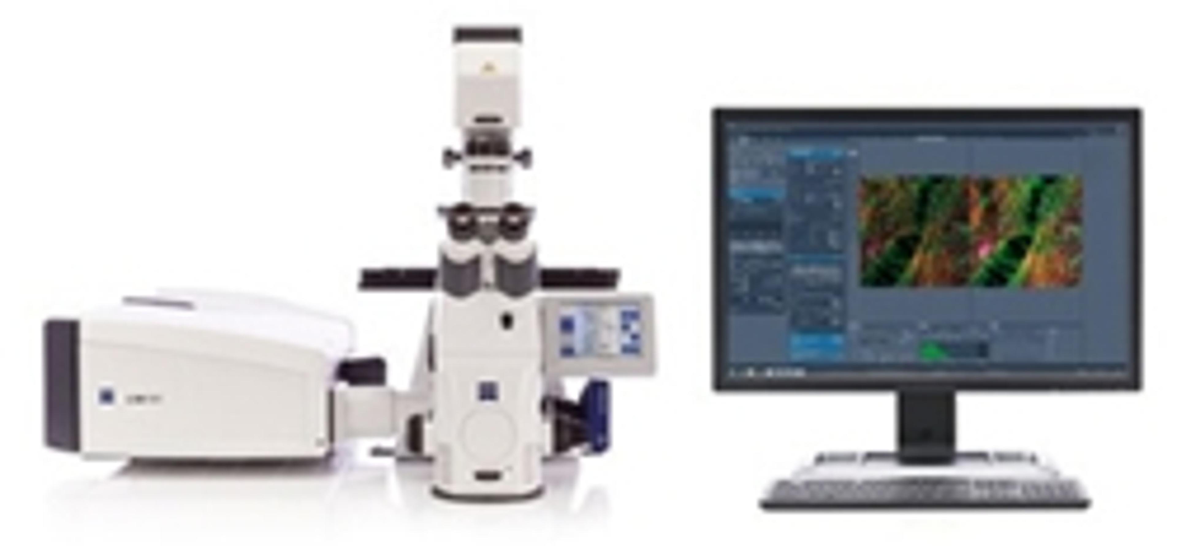
At the Annual Meeting of the Society for Neuroscience 2018 (Nov 03 – 07), ZEISS previewed new workflows in collaboration with Inscopix where ZEISS’s Airyscan confocal imaging technology was leveraged with the Inscopix freely behaving microscopy systems. The joint workflow development utilizes the increased spatial resolution, signal-to-noise and sectioning of ZEISS Airyscan to further inform the freely behaving signaling data collected with the Inscopix system. The presented workflow will serve as a preview of how the resultant data from the respective systems is correlated to benefit studies in functional neuroscience.
The benefits for scientists
Using Inscopix freely behaving microscopy systems allows researchers to monitor neural signaling during social interactions, addiction, sleep, spatial memory etc. Imaging depth is enhanced through the use of a GRIN lens enabling imaging from both cortical and deep brain areas. The new workflows previewed at Society for Neuroscience 2018 enable correlation of data collected in freely behaving animals with ZEISS Airyscan to provide additional contextual information by through optical sectioning, improved resolution, and improved signal-to-noise. In addition, leveraging the ZEISS Airyscan system allows for the potential of researchers to utilize additional fluorescence labels for structural context as well as potential to correlate with other multimodal imaging (i.e. X-ray microscopy, scanning electron microscopy/focused ion beam).
“The developments at Inscopix to enable co-registration of ZEISS Airyscan data with the large-scale neural activity data in freely moving subjects acquired with Inscopix's platform represents a long-sought marriage of two cornerstone imaging modalities. Integrating subcellular and structural detail with functional neural network data can provide exciting new insights into brain function and dysfunction,” summarized Dr. Kunal Ghosh, CEO of Inscopix.
“The vision is to see the strengths of the Inscopix approach and the ZEISS Airyscan technology combined in an overarching workflow makes us highly excited. We are convinced that joining forces with Inscopix to provide a solution integrating highly relevant functional and structural information is a perfect match”, says Bernhard Zimmermann, Head of the Business Sector Life Sciences at ZEISS Research Microscopy Solutions.
Discover more news from Neuroscience 2018 >>
The principle behind ZEISS Airyscan
A traditional confocal microscope scans the sample by illuminating one spot at a time to detect the emitted fluorescence signal. Out-of-focus emission light is rejected at a pinhole. Users can increase image resolution by making the pinhole smaller, but the collected signal drops significantly, since less valuable emission light is passing through.
With ZEISS Airyscan, ZEISS introduced a new detector concept. ZEISS Airyscan is a 32 element area detector. Each detector element functions as a single, very small pinhole, yet the overall assembly covers the area of a large pinhole. Knowing the spatial distribution of each of the 32 detectors results in improved resolution and signal-to-noise. The ZEISS Airyscan detector is available for ZEISS LSM 8 microscope systems and as an upgrade for the ZEISS LSM 710 and ZEISS LSM 780 confocal microscopes