Mapping the Unknown: How Mass Spec is Taking Us Deep Inside Tumors
Learn how innovative mass spectrometry techniques are enabling scientists to explore the molecular make-up of tumors — opening the way to better drug efficacy and new treatment strategies
20 Mar 2019
“One significant barrier for researchers is that we have an incomplete picture of the molecular make-up of tumors. With our project, for the first time, this picture is beginning to emerge.” Prof. Josephine Bunch’s exciting words show just how important the Rosetta project is in the fight against cancer. Bringing together experts from a range of fields, it aims to build a picture of a tumor’s microenvironment at a level of detail never seen before — a so-called ‘Google Earth of tumors’. Here, we catch up with Prof. Bunch, from the National Physical Laboratory, on the latest developments since we last spoke to her around 18 months ago — and the results so far, she tells us, are remarkable.

SS: Tell us about your project to map tumors using mass spec imaging
JB: The Rosetta project is developing a reproducible, standardized way of mapping tumors, at a level of detail that has not been seen before. By plotting single cells and the molecules inside them extremely precisely, we can draw a better picture of how a tumor changes and how it reacts to certain treatments, allowing us to predict how effective a treatment might be in the future.
It’s been described as a ‘Google Earth of tumors’ because, similar to these kinds of mapping services, which provide context and detail to help us to navigate the world around us, we want to be able to obtain context and detail in order to understand how a tumor changes on a molecular level. Just as Google Earth can zoom from a country down to a single street, we will be able to visualize whole tissues, and then have the capability to zoom into the desired level of detail, like molecules inside single cells.
We’re doing this by bringing together a range of mass spectrometry techniques, specialist instrumentation and methods at the National Centre of Excellence in Mass Spectrometry Imaging (NiCE-MSI) here at NPL, along with support from our collaborators at Imperial College London, AstraZeneca, Barts Cancer Institute and the University of Cambridge. We are using these methods to study cancer models from our cancer biology experts at The Francis Crick Institute, The Institute for Cancer Research, and the CRUK Beatson Institute. The group, formed of an international, multidisciplinary collaboration of chemists, physicists, and biologists was funded through Cancer Research UK’s Grand Challenge scheme — which helped to facilitate such an exciting mix of researchers coming together to take on this challenge.
Producing detailed information about the molecules in a cell, using fluorescent techniques for example, generally requires you to know what you’re looking for. Our work, on the other hand, is striding into the unknown. It’s genuine blue-sky research, but investigating this challenge is extremely important as, once we get this right, it could go on to make an extremely important difference. One of the ultimate goals of the project is new ways of measuring tumors. The better we understand the tumors, the better we can understand treatments, and can go on to design therapies. Just as the Rosetta stone was used as a reference point to decipher Egyptian hieroglyphics, our group is developing a reference point for identifying different tumors.
Currently, the Rosetta team focuses on a range of tumors, including breast, bowel and pancreatic tumors, as well as an aggressive type of brain tumor (glioblastoma). We picked these tumors as through our partners we have access to “state-of-the-art” models of cancer that recapitulate the human disease, as well as well-curated human samples, maximizing our opportunity to address major questions, for example around the mutational drivers within tumors.
The team are working to capture changes to these metabolites as tumors develop or respond to treatments, while simultaneously recording information about the cell’s exact location within the tumor, and over the past year has made impressive progress.
SS: Why is tumor mapping so important for cancer therapy?
We’ve made great progress with our understanding of some of the mutational drivers for cancer.
Prof. Josephine Bunch National Physical Laboratory
JB: Cancer is one of our most pressing public health concerns; it is widely reported that the disease is responsible for one in every four deaths in the UK[1]. One significant barrier for researchers is that we have an incomplete picture of the molecular make-up of tumors.
With our project, for the first time, this picture is beginning to emerge. Tiny changes in the metabolism of a tumor can have a massive impact on its overall state and function, and these changes can be observed using accurate scientific techniques.
SS: Why is mass spec so important for this project?
JB: Mass spectrometry is the ideal investigation tool. It separates the constituent parts of a sample according to their mass. It can tell you what is there, and how much of it there is. As such, we are able to identify metabolites — the molecules produced as a result of chemical reactions in a cell — and quantify them. This can be an indicator of the state of development and growth of a tumor cell.
We can generate vast amounts of data from these insights into metabolites, for example, about how tumors develop or respond to treatments, all alongside a precise measure of the location of the cell within the tumor.
We can then superimpose this data, which will all be made freely available to the research community, with maps that reveal information about the underlying genetics of the cells to generate high-resolution metabolic maps, offering unprecedented insight into the biochemistry of the cells to inform future developments in cancer therapies.
SS: What has the Rosetta project achieved to date?
JB: The project has progressed with impressive speed, and the results we’ve seen so far have been remarkable. In year one, we developed a pipeline for imaging tumors which produces an ultra-high-resolution picture of their metabolism. Working with our collaborators at Imperial College London: AstraZeneca, Barts Cancer Institute and the University of Cambridge, and cancer biology experts at The Francis Crick Institute, The Institute for Cancer Research and the CRUK Beatson Instute, we were able to validate this pipeline by analyzing pre-clinical tumors treated with different cancer drugs. This provided valuable insight into the distribution within the tumor and highlighted hot spots of drug resistance. By understanding the drug distribution within the tumor and linking this to biological effect, our mass spec imaging techniques are already enabling improvements to drug efficacy studies within the pharma industry.
We’ve also made great progress with our understanding of some of the mutational drivers for cancer and are ready to make the next step of seeing how pre-clinical model data will translate into human samples.

SS: How have Waters’ MALDI and DESI platforms been influential in this project?
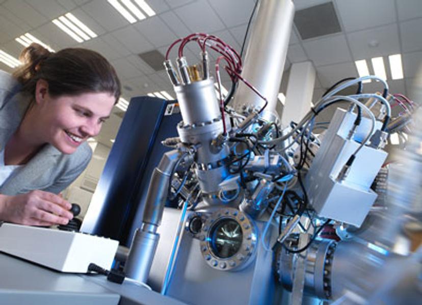
JB: The MALDI and DESI platforms both enable the spatially resolved detection of metabolites from surfaces of thin tissue sections. They are both label-free methods, which ionize (excite) the molecules from the surface either via a focused electrospray of a volatile solvent (DESI), or via assistance of a ‘matrix’ compound applied to the tissue, which is irradiated with a laser. The advantage of these methods, as well as being unlabeled, is that they are both relatively high-throughput methods that enable the rapid analysis of samples. They are suitable for analysis of different metabolites within the sample so are complementary to each other, and we are learning which platform is most useful for different experiments depending on the key metabolites of interest.
The real power of these platforms is that we can use them to identify and direct us to areas of interest (for example, areas of drug resistance) where we are then able to use much higher resolution methods, such as the labelled NanoSIMS, which offers the highest lateral resolution of mass spectrometry imaging techniques to image metabolites at an intracellular level of detail. Alternatively, we can use imaging mass cytometry which images mass tagged proteins with high special resolution. Both these high-resolution techniques have the potential to uncover an unprecedented level of detail within the tumor, but they are slow and expensive so it’s not realistic to use them in an untargeted fashion. Our pipeline enables us to harness their capabilities to better understand specific areas of interest.
SS: What impact will your findings have on cancer research and therapeutics?
JB: The first year of the project has already revealed a promising avenue for better understanding drug resistance, and this has been validated through our work with AstraZeneca; something that the whole project team are really excited about.
We are enabling our partners to begin to dissect and understand mutational drivers of cancer and how metabolism relates to activation of key pathways. In the long term, this will help us understand what the pre-clinical models are teaching us. We hope that ultimately this has the potential to bring forward new treatment strategies and biomarkers to develop them. We are definitely on the road towards our imaging pipeline identifying novel targets for drug development and providing accurate methods for diagnosis and monitoring treatment. We and our partners are excited to see if we can use these approaches for predicting therapeutic responses in these cancers.
[1] https://www.cancerresearchuk.org/health-professional/cancer-statistics/mortality
Do you use and Waters instruments in your lab? Write a review today for your chance to receive a $400 Amazon gift card>>

