Microfluidics technology provides physiologically relevant cell-based models for high-throughput drug discovery
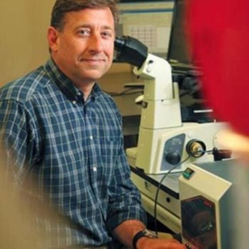
Jeff Jensen, CEO of Fluxion Biosciences, describes how microfluidics technology fuels advances in drug discovery through physiologically relevant cell-based assays
17 Oct 2023
Fluxion Biosciences is a leading manufacturer in microfluidic technology, enabling the use of physiologically relevant cell-based models for drug discovery and life sciences. Its innovative solutions benefit a broad range of research areas, including screening drugs that target ion channels and developing chimeric antigen receptor (CAR) T-cell therapy for solid tumors to name a few.
In vitro models are valuable tools for investigating response to disease and therapies. However, there is a need for more physiologically relevant cell-based models1,2 that more closely replicate response in the human body. Microfluidics, which can accurately model the in vivo environment, provides a proven technology for cell-based drug screening3.
Replicate human biology with microfluidics technology
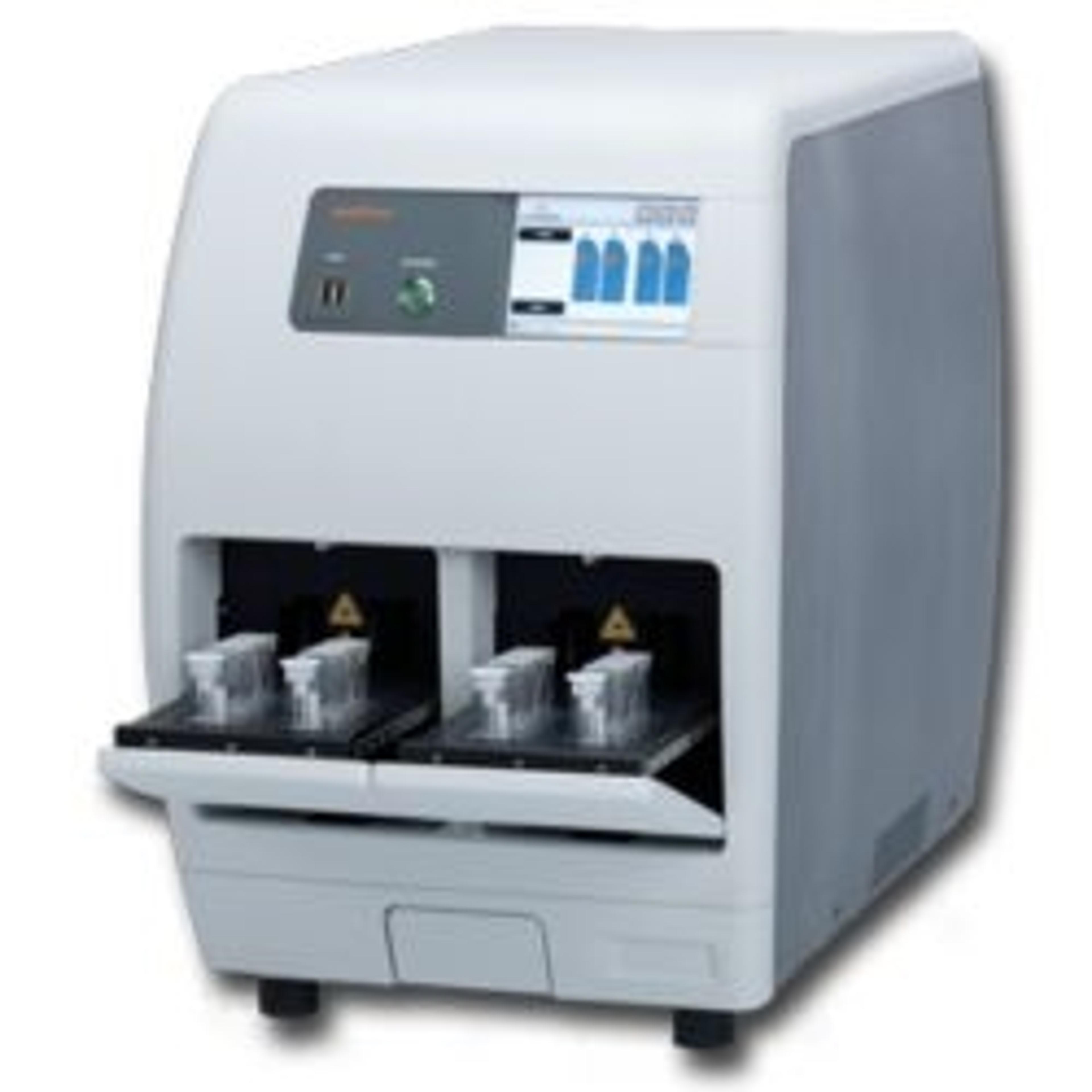
Fluxion Biosciences’ product offerings comprise three main families: automated patch clamp (IonFlux), cellular analysis (BioFlux), and liquid biopsy (IsoFlux). The underlying concept across all these product families is the ability to move, manipulate, and control the cellular micro-environment within microfluidic systems.
Microfluidics introduces a novel approach to studying drug-target interactions by allowing precise control of the cell environment. “The ultimate goal is to mimic cell behavior in the human body using microfluidic models, reducing the need for animal testing,” explains Jeff Jensen, CEO for Fluxion Biosciences. This aligns with the U.S. Food and Drug Administration (FDA) Modernization Act 2.0, which encourages alternative technologies to improve drug-target interactions using human-derived cells, making them more relevant and predictive than traditional animal models.
Jensen describes the core technology behind microfluidics, equating it to the development process of computer chips. “In essence, we apply computer chip manufacturing processes to create tiny silicon wafers with small channels, from which we mold our microfluidic ‘chips’,” states Jensen. “By leveraging this approach, we can shrink and multiplex experiments, performing hundreds simultaneously instead of one at a time. This scalability, coupled with precise control of the cellular microenvironments are the true advantages of microfluidics.”
A comprehensive microfluidics portfolio for diverse applications
Fluxion Biosciences' microfluidics technology has extensive applications across multiple fields with its IonFlux, BioFlux, and IsoFlux platforms. Applications span oncology, immunology, neuroscience, microbiology, and hematology to name a few.
“The IonFlux platform focuses on studying ion channels, which are crucial membrane proteins regulating various cellular functions,” Jensen explains. Ion channels serve as significant drug targets, but their modulation must be carefully controlled to avoid off-target effects, such as heart rate changes. IonFlux can be used to study the biology and pathophysiology of ion channels, enabling drug companies to identify drugs that effectively modulate ion channels, either inhibiting or enhancing their activity for different purposes; key focus areas for Fluxion’s IonFlux system include CNS disorders, molecular genetics, and cellular electrophysiology. Jensen adds, “this versatile platform is widely used from basic research in academic labs to high-throughput primary screening in the pharmaceutical industry.”
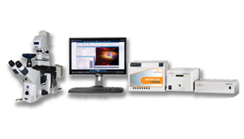
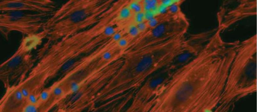
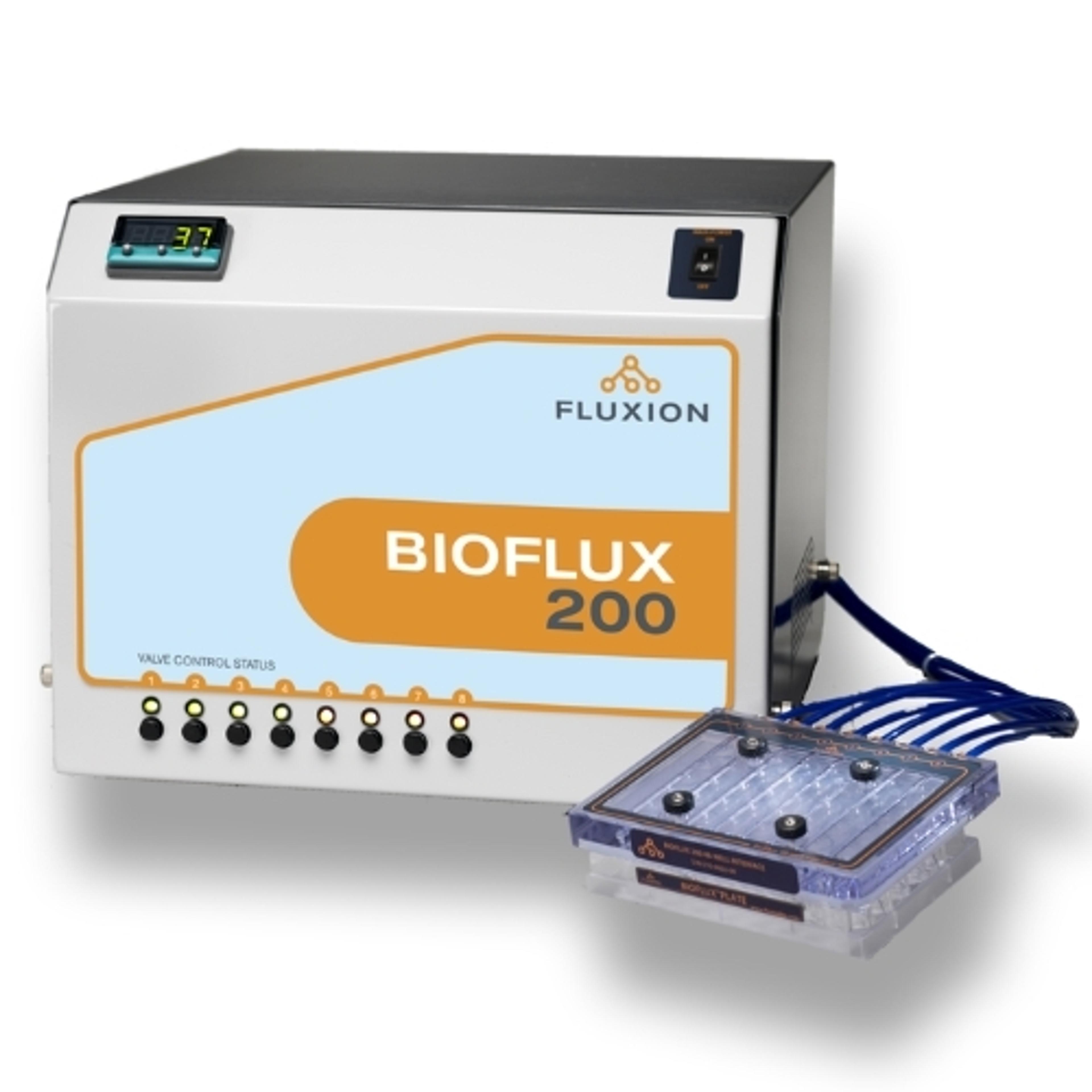
The BioFlux system is a platform tailored to studying cellular interactions under physiological shear flow. Jensen explains, “as an example, we can create an ‘artery on a chip’ where we replicate the conditions of an artery in the human body and study cell interactions within it”. The platform can utilize samples of blood directly, requiring only a small amount, typically around 100 or 200 microliters for each experiment. This is significant as it enables physiologically relevant research and the development of treatments for various conditions, including atherosclerosis, thrombosis, infectious diseases, and immune disorders.
“The third platform, IsoFlux, is for liquid biopsy and aims to enrich intact rare cells from biological samples and prepares them for further analysis, thereby providing a non-invasive way to diagnose cancer at the molecular level without performing a tissue biopsy,” says Jensen. “Instead, we analyze a patient's cancer using a blood sample through our IsoFlux platform.” This technology isolates and analyzes circulating tumor cells shed by the primary tumor in the bloodstream, providing molecular insights for accurate diagnosis and appropriate treatment decisions for the patient.
Harness microfluidic technology for physiologically relevant data
Many cells change their behavior in the presence of mechanical forces. For example, blood cells exhibit distinct behaviors when flowing through the circulatory system compared to being static in a well plate. “The dynamic flow causes the cells to undergo changes in their phenotypic behavior,” says Jensen. “Therefore, to accurately replicate the behavior of cells in the body, it is essential to have this flow capability included with the assay.”
To accurately replicate the behavior of cell in the body, it is essential to have this flow capability
Jeff Jensen CEO, Fluxion Biosciences
Historically, researchers created flow conditions for experiments on a macro scale by sandwiching together two pieces of glass or plastic to create a channel in the middle. Flowing blood or other cells through this could provide information on cell behavior and responses to potential therapeutic treatments under flow.
The traditional manual system was laborious and low throughput, so Fluxion Biosciences took this capability and integrated it into a microfluidic approach, streamlining the workflow and increasing the throughput. This newfound ability to run many experiments simultaneously in this miniaturized format has great significance in drug discovery, changing the way scientists can carry out cell interaction studies for a broad range of applications.
BioFlux finds diverse applications in immunology, cancer, CAR T therapy development, infectious diseases, sickle cell disease studies, and various other areas. Researchers utilize it to gain functional insights, evaluating the performance of candidate drugs and understanding their efficacy.