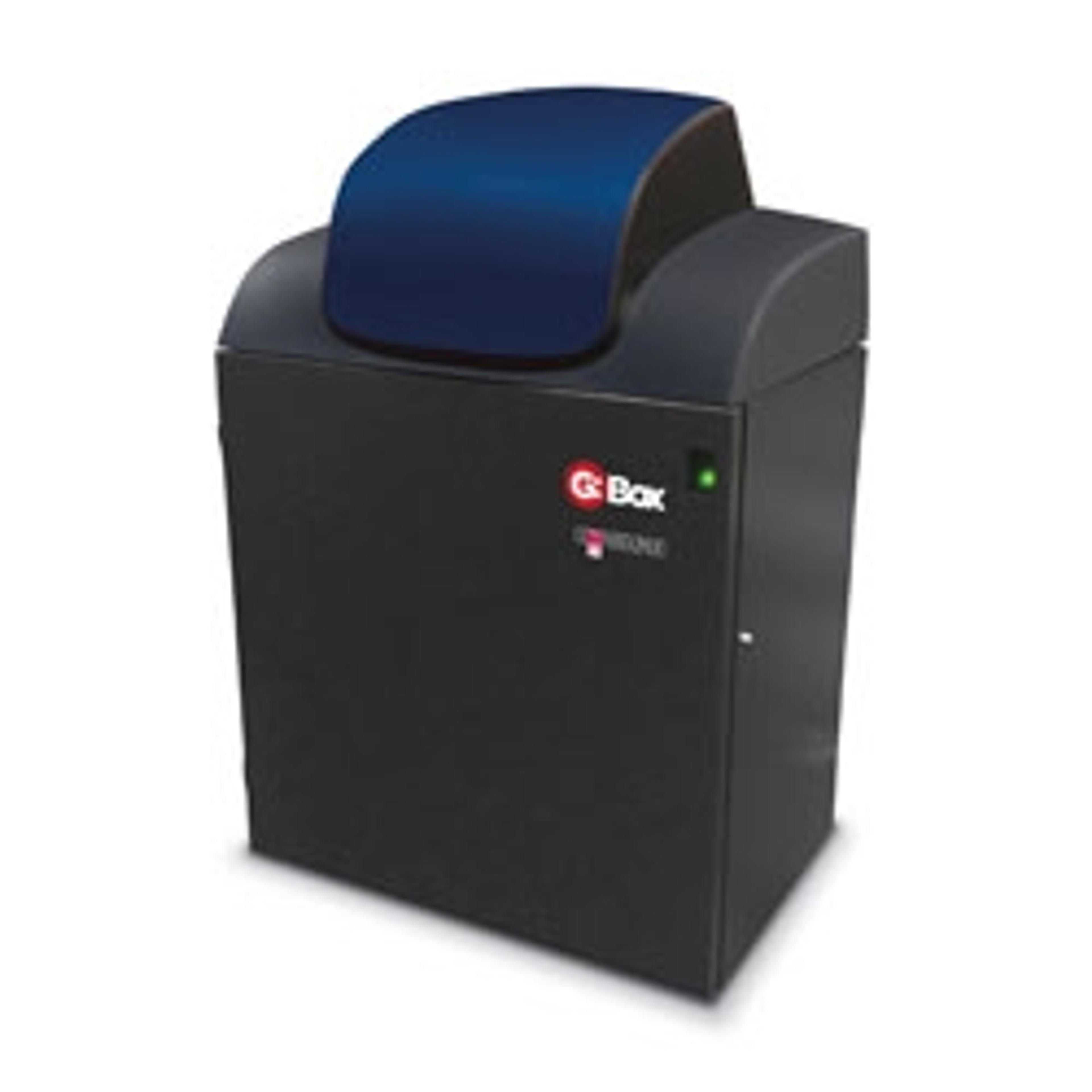
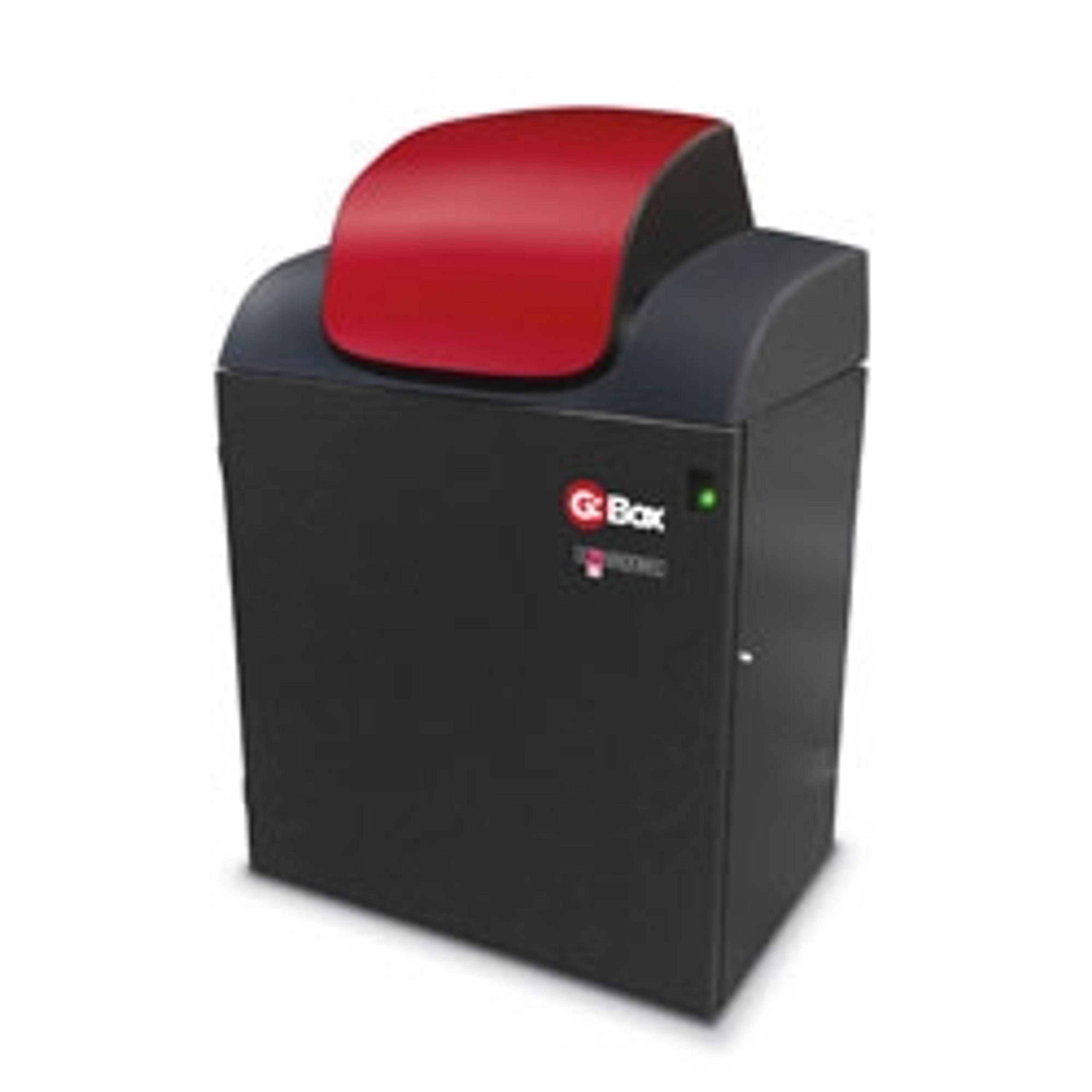
New IR Imaging Application of Syngene G:BOX Chemi Systems
23 Feb 2011Syngene announced that its new range of G:BOX Chemi advanced multi-application image analysers can be used for imaging with infra red (IR) LI-COR IRDye® dyes, making it easier to detect and quantify different types of proteins on multiplex Western blots.
The G:BOX Chemi systems, when fitted with a combination of recommended lighting and specific Syngene filters can be used for imaging LI-COR dyes, IRDye® 680 (Epi Red Multiplex LED lighting module and Syngene 705M filter) and IRDye® 800 (Epi LED IR 740 lighting module and Syngene LY800 filter). The GeneSys software in the G:BOX Chemi automatically selects the right lighting and filters for whichever IR dye or other fluorescent dyes scientists inform the system is on the blot. The software then captures one perfect image of all the different dyes, to ensure imaging fluorescent multiplex Westerns is quick and simple.
Laura Sullivan, Syngene’s Divisional Manager explained: “Scientists want to use fluorescence to visualise proteins on Western blots because they can increase throughput by using the same blot to detect different proteins, something they can’t do using chemiluminescent-based blots. Additionally, it can often be difficult to accurately quantify proteins as some fluorescent dyes have overlapping spectra and membranes can auto-fluoresce, which interferes with detecting low abundance proteins. Using IR dyes can sometimes solve these problems.”
“Detecting IR dyes has proved difficult using CCD-based systems and so we are excited to have found filters and lighting combinations to allow our G:BOX Chemi to visualise multiplex Westerns of LI-COR IR dyes. This breakthrough means scientists with a G:BOX Chemi now have a sensitive, accurate method for imaging IR labelled proteins without having to buy an expensive laser-based scanner,” Laura Sullivan added.


