New Leica imagers set to transform visualization of 3D samples
The THUNDER imager family enables users to decode 3D biology in real-time*
23 Jan 2019
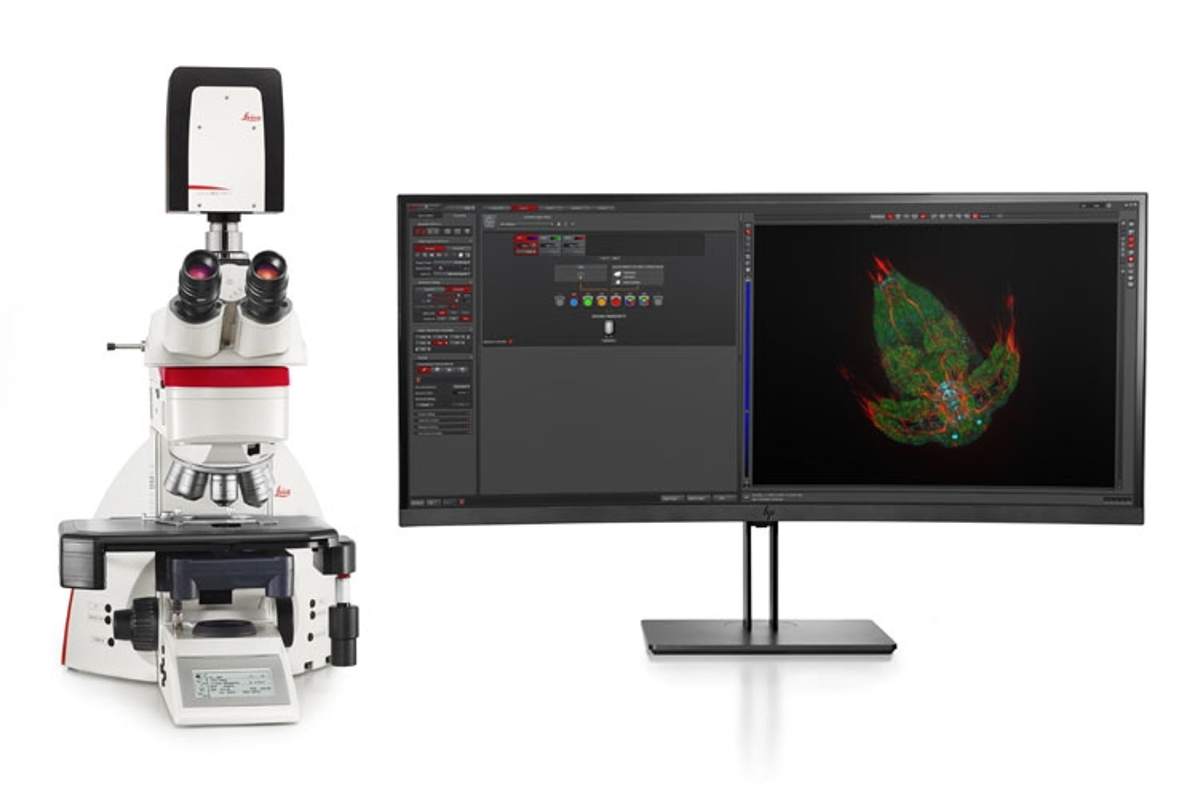
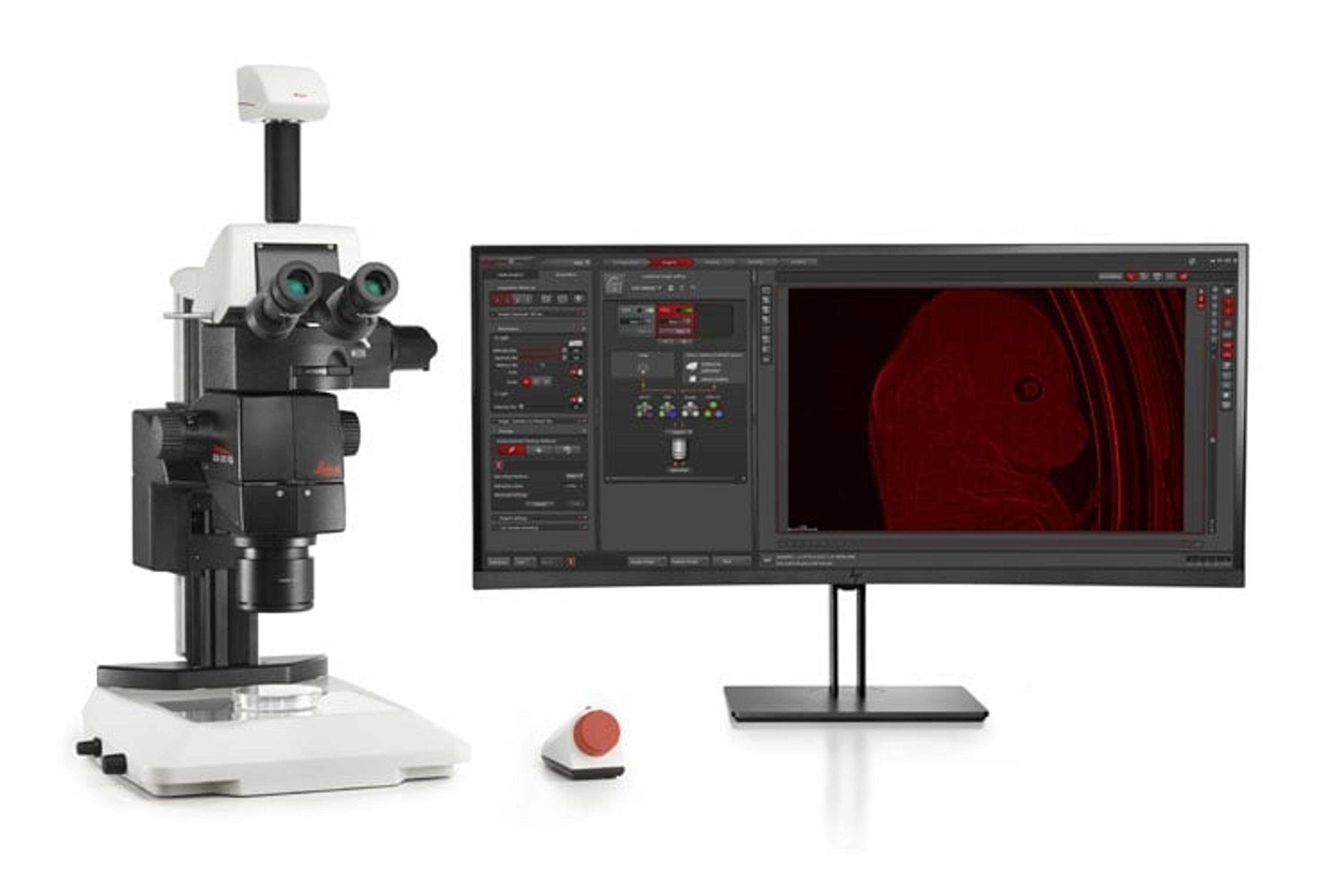
Leica Microsystems, a world-leading designer and manufacturer of innovative microscope solutions, has announced the launch of a new class of instruments for high-speed, high-quality imaging of a large diversity of samples, including thick specimens. THUNDER Imagers allow to visualize clearly in real time details even deep inside thick samples of biologically relevant models like model organisms, tissue sections, and 3D cell cultures. This makes THUNDER Imager a cost-effective and speedy alternative to methods like structured illumination or spinning disc.
THUNDER Imager eliminates the out-of-focus blur that clouds the view of thick samples when using camera-based fluorescence microscopes. This performance advantage is achieved with a new opto-digital method created by Leica Microsystems called Computational Clearing. Currently unique to the market, the new technology deployed in THUNDER Imagers makes visualization and analysis of large volume, thick specimens ideal for many biomedical applications where they are required, including regenerative medicine, cancer, and stem cell research to decode 3D biology in real time.
Markus Lusser, President of Leica Microsystems: “The innovations we are investing in at Leica Microsystems are all about maximizing output and reducing cost and complexity, leveraging our 170 years of expertise. We are extremely proud of this new family of instruments we are bringing to market, which will help our customers meet challenges in the laboratory that come with the trend towards using always more biologically relevant specimens that are typically thicker.”
The new series of THUNDER Imagers are available in configurations for three application areas:
- THUNDER Imager for 3D Cell Culture & 3D Live Cell are designed for imaging of cell culture assays. It helps to maintain optimal physiological conditions by minimizing photobleaching, providing high-performance imaging and high-throughput of data leading to better workflow efficiency and statistics.
- THUNDER Imager for Tissue is designed for real-time 3D fluorescent imaging of thick tissue sections. Typically used for neuroscience and histology research, this system combines the speed, fluorescence-signal sensitivity and ease-of-use common to widefield microscopes, giving access of a tissue's finest structures even deeper in the sample
- THUNDER Imager for Model Organisms enables imaging of organisms used for developmental or molecular biology research, such as Drosophila, C. elegans, zebrafish, mice, etc. It delivers blur-free images revealing the fine structural details of live organisms, while keeping them under optimal physiological conditions.
* in accordance with ISO/IEC 2382:2015
Want the latest science news straight to your inbox? Become a SelectScience member for free today>>


