Novel methods for the development of stem cell-derived 2D and 3D models
Watch this on-demand webinar to explore the use of hPSC-derived models to study human health, development, and disease
25 Jul 2023
Stem cells are an invaluable tool for generating multiple cell types from individual patients. However, the workflows associated with growing, differentiating, and CRISPR gene editing induced pluripotent stem cells (iPSCs) in 2D and 3D culture are inefficient, low-throughput, costly, time-consuming, and manually labor-intensive.
Human pluripotent stem cell (hPSC)-derived organoids are providing unprecedented access to study health and disease in human-specific model systems. For example, choroid plexus organoids are a convenient in vitro system for studying the brain’s blood-cerebrospinal fluid (CSF) barrier and the production of CSF, which can potentially reduce or supplement the use of animals in research.
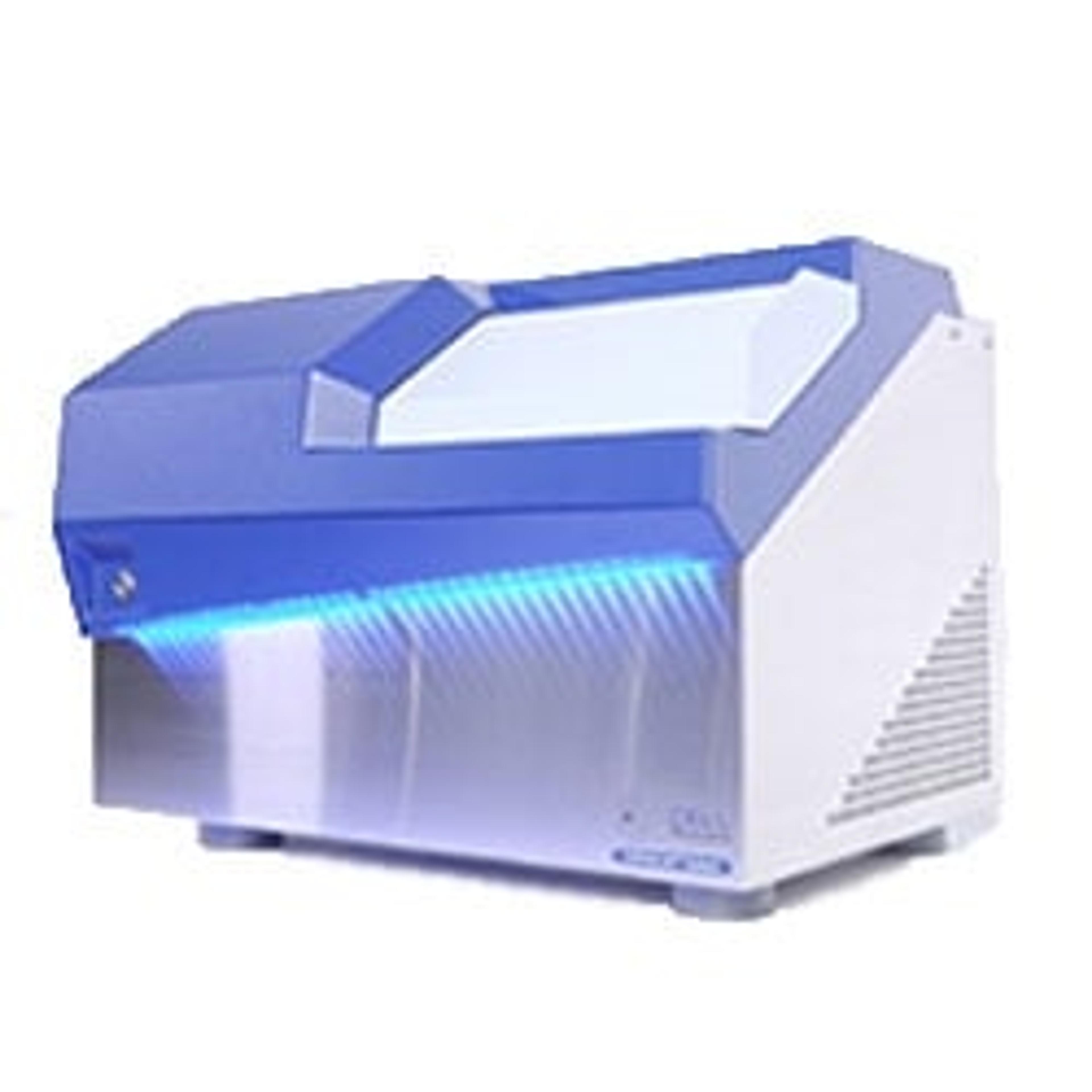
In this webinar, now available on demand, STEMCELL's Dr. Erin Knock describes how to culture and differentiate choroid plexus organoids derived from hPSCs, and explains how they can be applied to answer specific research questions. Dr. Jessica Hartman from Cell Microsystems discusses improving workflows that span the breadth of iPSC biology, from reprogramming to monoclonal edited colony formation to 3D models for disease. She also provides an overview of CellRaft technology, which includes the CellRaft® Array, CellRaft AIR® System, and CellRaft Cytometry™ software.
Watch on demandFind highlights from the live Q&A session below or register to watch the webinar at any time that suits you>>
Can the software be used for any type of stem cells, or is it made for iPSCs specifically?
JH: The software is adaptable to any cell type that you want to use on the CellRaft AIR System – that's really the power of the software, and why it's such a great tool. Because all the characteristics are totally user-defined, you can use it to set any characteristics that are important to you for any cell type that you're using. It also makes the AIR system very versatile, because as I mentioned, we've used it for over 80 different cell types ranging from adherent to suspension to primary, and you can create custom parameters for each of those. So yes, it's adaptable for any cell line that you're growing in your lab.
Do you think this technology would be useful in looking at drug treatment in stem cells, or is this more targeted towards cell line growth and differentiation?
JH: It could definitely be adapted for a drug treatment or response. We've had some customers do those workflows before, directly on the array. So, if you're looking at drug sensitivity or resistance, you can do that on the platform as a cell-based assay, and isolate clones that respond one way or the other, linking phenotype to genotype. Alternatively, you can create a custom 2D array of your cells just like you would with the 3D arrays that I talked about. So, if you were doing drug screening on a broader level, and you wanted to put an established iPS cell line into a 96-well plate to do multi-time point or multi-dose type drug discovery, that would be possible as well.
You mentioned being able to separate the cells to observe phenotype. Would we be able to extract the cells from the dish to see genotype changes?
JH: Yes, you can do that one of two ways. You can isolate directly off the CellRaft Array into a lysis buffer. So, rather than isolating into a 96-well collection plate, you can isolate into a PCR plate or a PCR strip tube into lysis buffer. If your endpoint is lytic, you can isolate into a lysis buffer or another type of assay buffer. If you're looking for clonal expansion, or you've edited, and you're trying to look at the genotype downstream, you can always let your clones grow in the 96-well plate. Then, once they've grown off the raft and they've attached to the floor of the 96-well plate, you can do whatever you like with them downstream. It's very adaptable to other technologies that you might have in your lab, whether that be a library prep system or a plate reader, for example. You can certainly use the clones that you generate to do any other downstream analysis that suits your workflow.
Is there any possibility of cross-contamination using the same magnet bar?
JH: We get this question a lot. The possibility of this is really, really slim. The cells stay very tightly attached to the Raft, just like they do to any other polystyrene growth surface. Also, the transfer time on the wand is quite low, so, the dwell time for the Raft is about five seconds from pickup to touchdown. So, the possibility of transfer is really slim. We've also tested this extensively, using what I call a checkerboard experiment, where we isolate different phenotypes of cells into collection plates in sort of a mixed pattern, and we don't see any cross-contamination during that transfer period. So, the likelihood is really very low.
Watch this webinar on demand here>>
SelectScience runs 10+ webinars a month across various scientific topics, discover more of our upcoming webinars>>

