Nucl:Cyto Segmentation in HCS Imaging Enabled by Far-Red Dye
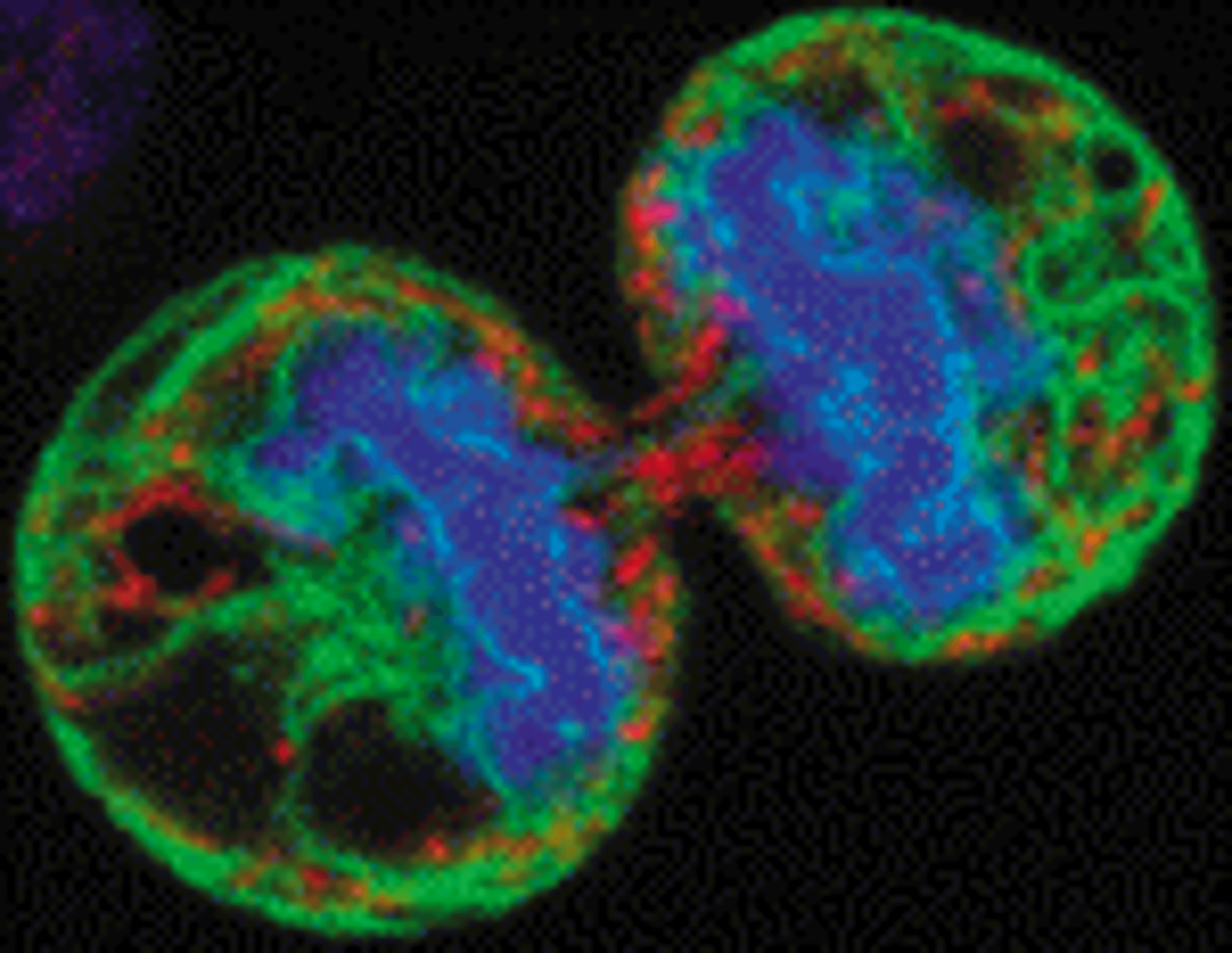
24 Jun 2008Scientific presentations at the recent HCA Europe conference in Prague confirm the nucleic acid-binding dye DRAQ5™ as counterstain of choice for cell-based imaging HCS assays. DRAQ5™ rapidly enters live or fixed cells to intensity discriminate between nuclear and cytoplasmic compartments. The resulting differential is ideal for compartment segmentation by automated image analysis tools. This offers new opportunities for cell-based assays where cell location, cell perimeter, cell shape and cell spread parameters can be used to define the assay at the single cell. DRAQ5™ permits the reliable tracking of fluorophore-tagged protein translocations and expression changes in GPCRs, kinases and other therapeutic targets in drug discovery and in vitro toxicology.
The far-red, nucleic acid binding fluorophore DRAQ5™ avoids emission overlap with most visible range fluorophores and importantly GFP and related tags. This simplifies assay development, making it more flexible, enabling multiplexing and the standardization of one key image-based assay parameter. DRAQ5™ is compatible with all HCS cell imaging platforms including Opera, IN Cell, ImageXpress and ArrayScan.
DRAQ5™ assays use one fluorophore to reveal both major cell compartments. Acquisition of both these and the target protein signals can be done simultaneously, offering a halving of plate scanning times. UV excitation is avoided reducing interference from compound auto- and cellular bio-fluorescence.
Ready to use, DRAQ5™ rapidly labels live or fixed cells with little need for optimization. Staining is stable and insensitive to MDR phenotypes. The simple, no-wash procedure is already widely used in temporo-spatial, fixed end-point and homogeneous assays across drug discovery. DRAQ5™ is both chemically stable and resistant to photo-bleaching. It is compatible with aqueous media and may be combined with cell fixative to remove a protocol step. DRAQ5™ additionally, provides DNA content information as recently demonstrated on the Acumen eX3 microplate cytometry platform.
Biostatus offers bespoke presentation of DRAQ5™ to suit the needs of its high-volume HCS users to further streamline assay protocols and increase workflow efficiency. These data confirm that DRAQ5™ improves HCS assays - technically, operationally and financially.

