On-Demand Webinar Highlights: Understanding Cell Motility Through Next-Generation Scratch Wound Healing Assays
Learn how to get the most from your scratch wound healing assays
26 Mar 2018
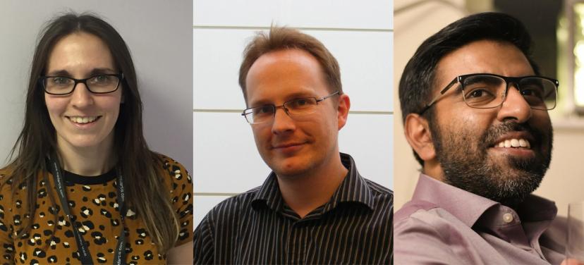
In our on-demand webinar , Dr. Rakesh Suman, senior biological application scientist at Phasefocus, discusses how to significantly enhance conventional wound healing assays by diminishing common assay limitations, and introduces the Livecyte™ Kinetic Cytometer that is enabling these advances in live cell analysis.
Providing research examples and advice on in vitro experimental techniques, Dr. Mat Hardman and postdoctoral researcher, Dr. Liz Roberts, also discuss how the Livecyte system has helped to propel their research.

This webinar explores:
- How to separate cell motility from cell proliferation
- The advantages of continuous monitoring of the wound healing process using label-free Quantitative Phase Imaging (QPI) imaging
- The ability to segment and track individual cells along the leading edge to directly measure cell motility
- Characterization of morphological and dynamic cell phenotypes during wound healing
Watch this webinar on demand >>
Q&A highlights
Q: What’s the longest time-period that you have imaged cells? Were they still alive at the end?
Rakesh. On the Livecyte, we’ve imaged primary hippocampal neurons, so very sensitive primary cells. With that cell type, we could image for seven complete days with an imaging frequency of every six minutes. In theory, you can keep your cells alive and image for weeks using the Livecyte system, as long as you incorporate a media change.
Q: Is there a difference between scratch wound and exclusion assays for measuring cell migration, and which is best?
Mat. It all depends on what outcome you’re looking for and what question you’re asking. If you make a scratch wound, you’re injuring the cells, so you have an added component of injury and cell migration. Whereas in an exclusion assay, if you’re excluding an area and then looking at the cells migrating into that, the cells migrate much more quickly and don’t have an injury component. So, I don’t think that one’s better than the other, but they’re two different ways of answering two different questions.
Q: What’s the best way to create a uniform and consistent wound on the plate?
Liz and Mat. Firstly, it takes a lot of experience! The more scratches you do, the better you get at making them more uniform. In terms of making the scratch:
- Try and keep your pipette tip as perpendicular to the well as possible, so that you’re not going in at an angle.
- Confluence is important. You need uniform cell confluence throughout the well. Too confluent and the cells will start to peel off when you’re making the scratch.
- Use the edge of the plate as a guide to try and make the scratches as straight and uniform as possible.
- Pressure is important. You need to try and get an equivalent pressure all the way down the well.
- In the exclusion assays, you can seed directly into inserts. When you remove your inserts, you’re left with a very uniform gap between your wells.
- In the Livecyte system, you get to see your scratch at your time zero point, immediately after you’ve made the scratch. This helps you spot any disparity between the wells at the very beginning, which you don’t see in your end-point assays.
Watch this webinar on demand >>
Q: What type of cells were you working with for the scratch wound assay, and can we define scar formation?
Rakesh. We’ve tried a lot of different cells during research at Phasefocus. The cells shown in the slides were MDA-231 metastatic breast cancer cells. Due to the negligible phototoxic risk to the cells when using the Livecyte system, some of our users have also been able to work with primary cells.
Mat. A scratch assay is not really applicable in terms of measuring scar formation, which is essentially matrix deposition and turnover. However, you can culture cells on matrix for a scratch assay. So, you can potentially culture cells on matrix that has come from primary cells isolated from a scar-prone situation versus a non-scar-prone situation. The other aspect of scarring is fibroblast differentiation, so you could potentially use the Livecyte to quantify parameters that are linked to differentiation of fibroblasts into a more scarring phenotype.
Q: What is the percentage of wound closure being quantified?
Rakesh. We quantify the percentage of wound closure by taking an initial area of the wound and then, at each time point, we express what the area is at that point as a percentage of the original wound area. As the wound heals, it will reduce from 100%, all the way down to 0% as the wound closes.
Mat. As well as basic scratch wound closure measurements, the Livecyte system also brings a completely different dimension to this assay, in that it can measure the behavior of individual cells within a scratch. This allows you to do things you can’t do with other conventional methods and look at how individual cells are responding alongside how the scratch cell population is closing as a whole.
Q: What is the optical performance of cells embedded inside a 3D gel, such as a collagen gel?
Rakesh. You can image cells seeded on top of gels, but the performance depends on the quality and optical clarity of the gel. We find that a thin layer of collagen or Matrigel is fine to image through.
Q: What is the significance of being able to track leader cells and the cells further back in the scratch wound? Why is this important?
Mat. A lot of the time when people look at scratch migration, particularly in epithelial cells, you tend to focus on how fast the front of cells are moving to close the scratch. But that’s only one aspect of the biology of how cells move and how cells close. When you injure an epithelial front, you get a wave that moves away from the front edge of the scratch that tells the cells further back that they need to become activated and start migrating to be able to close the scratch. The cells need to change their cell adhesion and phenotype to allow them to close the scratch. So, if you can start to break the front down into leading edge cells, and the rows going back, you can start to understand how the cells are changing their phenotype in a collective manner.
Q: What is the biggest field of view that you can image and does it always have to be square?
Rakesh. On the Livecyte, the biggest area that you can image is 2 x 2mm (2000 x 2000 microns). The field of view doesn’t have to be square all the time. We can edit that to be letterbox in both portrait or landscape mode, with longest length being 2mm. We can drop multiple fields of view in a well, so you can have as many as you want across the whole leading edge if you wish to do so.
Q: Based on your knowledge, which cell line is preferable for wound healing assays?
Mat. It depends on what you want to look at. A scratch assay is probably best to look at epithelial cells if you want to look at epithelial cell collective migration. But if you want to mimic keratinocyte function to close a wound in the skin for example, then you’re best looking at primary keratinocytes to give you the most biologically-meaningful information. But you can use a cell line to mimic the effects of keratinocytes. It all depends on the questions you’re trying to answer.
Learn more about wound healing techniques: Watch this webinar on demand >
