Optimizing cryopreserved tissue samples using cell sorting to study cellular heterogeneity
Watch this on-demand webinar to learn how the dissociation and cryopreservation of primary solid tissues can accelerate preclinical pipelines
23 Nov 2021

The generation of a highly annotated cryopreserved dissociated tissue biobank provides the opportunity to develop highly focused study cohorts to understand the complex cellular heterogeneity of human tissues. Performing omics analyses on heterogeneous biological tissue samples can, however, lead to biased results, particularly when the target cells are present in relatively low numbers.
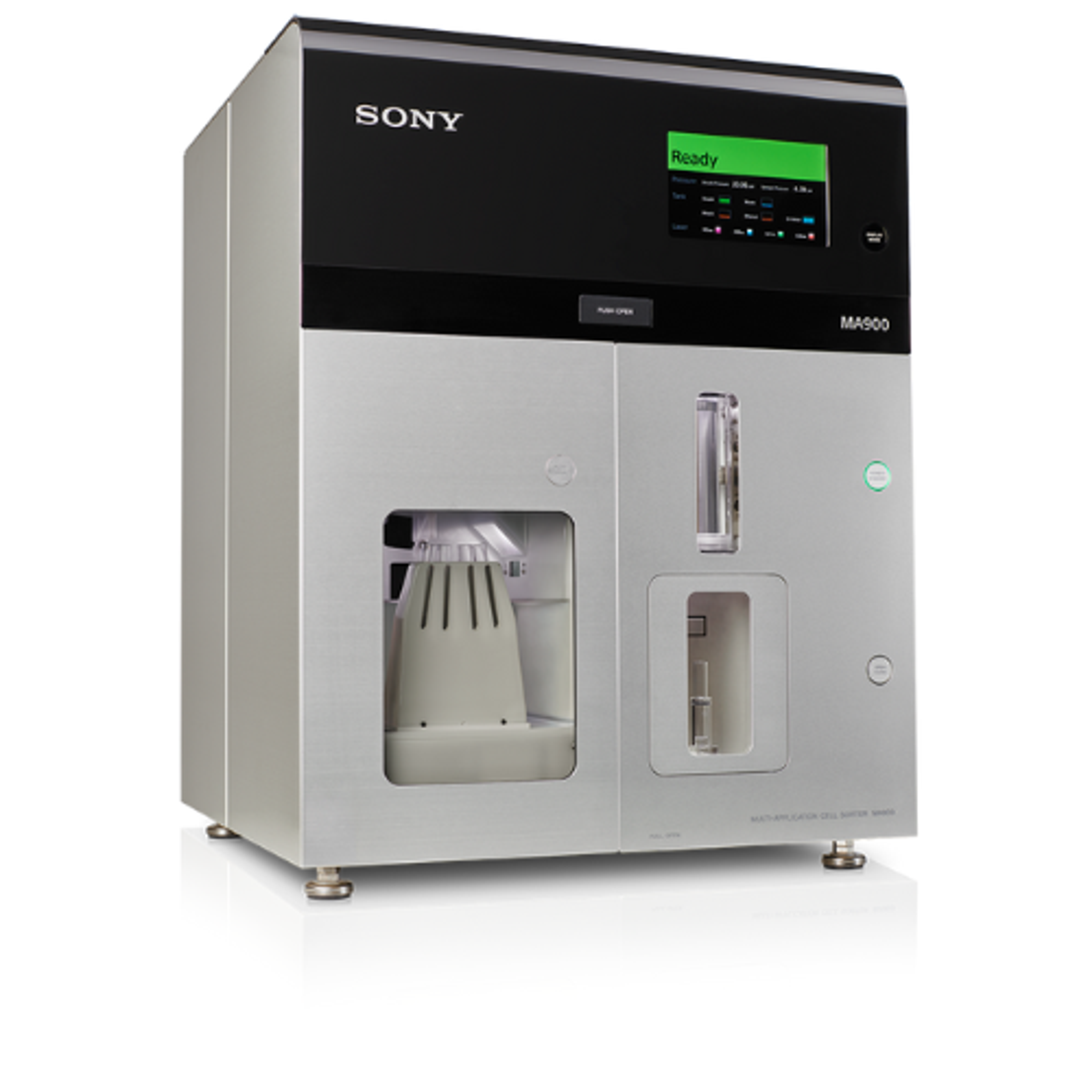
In this free SelectScience® webinar, now available on demand, Dr. Shawn Fahl, senior director of R&D at Discovery Life Sciences, describes how primary human tissue is processed for downstream analytical evaluation within Discovery Life Science’s cell services laboratory. Plus, Fahl introduces Sony’s MA900 cell sorter, and provides experimental data showing single-cell resolution of protein expression and results using 10x Genomics’ single-cell RNA-seq (scRNA-seq) technology.
Watch on demandRead on for highlights from the Q&A discussion and find out how Sony’s MA900 cell sorter is an integral part of multiple workflows that support both the analyses of cell populations and preparation of specimens for multiple downstream applications.
Do you have any capabilities for single nuclei RNA sequencing using frozen tissues?
SF: The way that these studies have worked is, for every frozen type of tissue, if it's from a different organ or a different tumor type, we really optimize how to get those nuclei out. We tend to vary the time of the nuclei isolation as well as the concentration of the buffers to make sure that we're getting an efficient release of those nuclei. Then, the key is the cell sorting. When we do this, only about 10% to 20% of the material that the cell sorter has seen are nuclei, by either a DAPI or a 7-AAD stain.
It's become crucial for us to identify these. Hopefully, very soon, we'll have a nice data set that we can start sharing to show how we've taken frozen tissue into that pipeline.
Do you work with dissociated tumor cells (DTCs) from normal adjacent tissue?
SF: This is something we've been trying to do for quite a while. When the tissue is resected, the margins are generally very minor. This is great for the patient, and usually, any of that normal adjacent tissue is embedded for the pathologists. Most of what we do receive is in embedded formalin-fixed paraffin-embedded (FFPE) blocks. So, that's where we've moved more to cadaveric donors for most of our normal tissue, just because that is a more readily available option than normal adjacent tissue.
How does RNA sequencing compare between samples from FFPE and fresh tissue?
SF: This is something that we've just started to take on in a couple of different formats. We have a large study where we're getting renal tissue in, and that renal tissue is matched with an FFPE block that's embedded inside. That tissue comes in, we dissociate internally, and then we test directly after dissociation into the 10x platform, cryopreserving that sample, having to go through a freeze-thaw, then that goes to the 10x platform.
We've also just started the VisiumTM Spatial Transcriptomics platform internally as well. So, eventually, we'll have a data set, which will show us both the fresh versus fixed RNA profiles, as well as fresh versus frozen to really understand what's the best specimen for a specific study. While we fully anticipate that there will be some subsets that are negatively impacted either by fixation or freezing, there are some that will not be. We're going to use this as a data set to help scientists choose the specimen types that they need for downstream studies.
When using cell sorting methods to separate debris from DTCs, how do you limit any further loss of live cells?
SF: This is where the cell sorter that we chose to use is crucial. We picked the Sony cell sorter because it gives us a lot of ability to manipulate the pressure of the sample going through the line. Most flow cytometers, cell sorters, have three options of a low, medium, high.
The Sony cell sorter goes from 1 to 10, or even from 0 to 10, so it gives you a lot of ability to bring that pressure down low. We tend to run these at fairly low pressure, and very low event rates to get minimal cell shearing through the system. We also then double-check at the end with a second cell count before we go in. Luckily, with 10x, we don't need a huge amount of specimen to go into the system, so we try to get the most high-quality system even though the yield might be a little bit lower, but we know that it's the most live it can be going into the system.
Do you ever lyse red blood cells prior to analyzing tumor samples by flow?
SF: Originally, we tried this and got worried. Given how much this tissue has to go through the dissociation process, we didn't want to stress it out even more through a red cell lysis step. What we do instead is every panel we run, we throw in a marker that marks red blood cells. If it's a frozen sample, there tends to not be many red blood cells remaining. If it is a fresh sample, though, there is a ton of red cells remaining in every indication we've looked at. We use CD235a to gate those out. It helps clean up the sample when you're doing flow.
With matched dissociated cells and FFPE blocks, have you or are you planning to implement spatial transcriptomic platforms?
SF: That's our biggest new pipeline that we're going to be starting. We just got the microscope in, which is needed to do the scanning of the slides. We anticipate having VisiumTM specifically because we have a good relationship with 10x. We expect that to be up and running by the end of the year, so that will open up both the DTC versus FFPE as well as just an FFPE spatial transcriptomics offering.
Do you think that magnetic bead-based dead cell removal could help here?
SF: It can. We have tried a couple of systems based on the magnetic bead from Miltenyi and STEM, and they do work. Sometimes you get a loss of a lot of cells through this process, so while it comes out very clean, your yield is very low. We haven't quite figured out why yet, but a lot of it has to do with the amount of Annexin V going into the system. We're having to tighten that down to make sure we're not selecting cells that might be alive and may become apoptotic, so have a low-level expression. I know 10x likes to use this system when doing cleanups. We've veered away from it as we have the ability of the cell sorter here. We've got the pipeline working well, so, because of that large loss of cells, we focus primarily on cell sorting.
What are the viable storage times for the samples that you've been using?
SF: In terms of the fresh samples coming in, we try to receive as many as we can within 24 hours. If these are coming from overseas into our Alabama location, or even in the European location crossing some of the country borders, that can be extended a little bit. So, we've got some good data that shows that up to about 72 hours after resection in the tissue storage solution we used, the samples are still very viable.
Part of the study that we were doing with the fresh versus frozen versus FFPE, was taking the tissue when it comes in and splitting it in half. Half gets processed immediately, so within 24 hours, and the other half sits until 72. So, there'll be an additional data point on top of that that will also look at time from resection to processing and then how cryopreservation affects both of those. We're hoping to have that data set, again, in the near future.
Can you discuss any studies you've performed on the differences between fresh and cryopreserved dissociated samples?
SF: We've done a fair amount of flow. The one subset that seems to be the most sensitive is the CD15-positive cells. These tend to be your granule acidic populations, so not entirely surprising that those don't survive the freeze-thaw. That’s not to say that they're not there, we do have some samples that after the freeze-thaw have a ton of CD15-positive cells, but they tend to be the most sensitive.
We did do an additional study that looks specifically within T cells, but these were T cells fresh from dissociation versus frozen and we looked at repertoire analysis and RNA-seq. We overlaid the fresh versus frozen, and they were virtually identical. So, while something like a granule acidic cell seems to be a little more sensitive to the freeze-thaw, the T cells, at least from the RNA and the TCR levels, seem to be fairly consistent.
Learn more about utilizing cryopreserved primary solid tissues: Watch this webinar on demand>>
SelectScience runs 10+ webinars a month across various scientific topics, discover more of our upcoming webinars>>