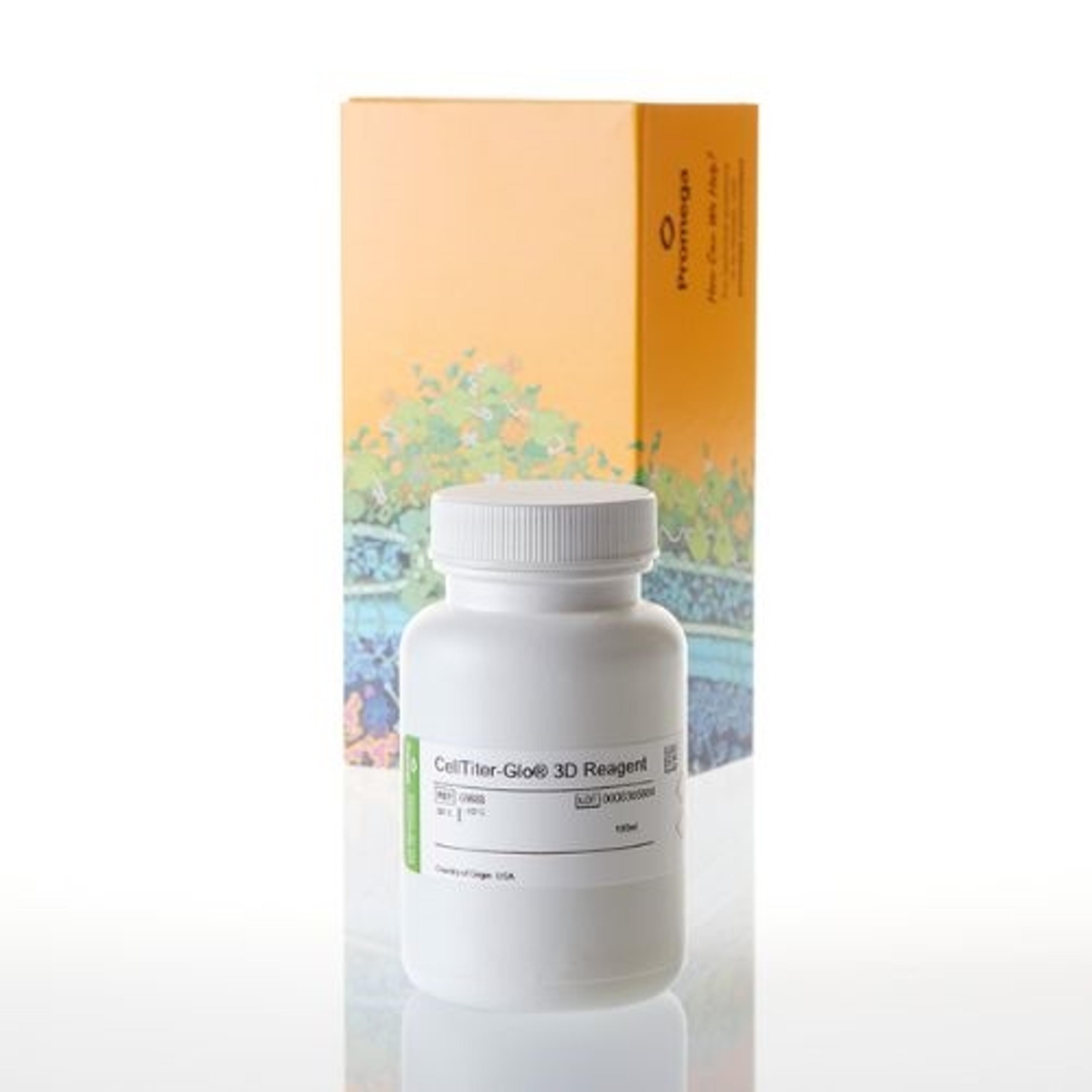
Promega Cell Viability Assay Designed Specifically for 3D Microtissue Culture
11 Jun 2014
Promega Corporation launches a new assay validated for assessing the viability of cells in three-dimensional (3D) microtissue model systems. CellTiter-Glo® 3D Cell Viability Assay penetrates large 3D microtissue samples to provide more accurate determination of viable cells. The assay has been validated on a range of 3D microtissue model systems, generated using scaffold and scaffold-free systems. CellTiter-Glo 3D Assay uses the robust Ultra-Glo™ Recombinant Luciferase in a solution optimized to penetrate denser 3D microtissues for a complete release of ATP and accurate assessment of viability by luminescence detection.
Scientists have found that 3D microtissue cell culture models better reproduce the cell-to-cell and cell-to-matrix interactions occurring in living organisms, providing a more predictive in vitro experimental model system. Most, if not all commercial cell-based assays, including MTT, resazurin and current ATP assays, were developed using 2D monolayer models, are less effective on denser 3D microtissue models and cannot fully penetrate larger 3D microtissue spheroids.
"There is a clear need for cell viability assays validated for use in 3D microtissue culture. The novel chemistry of CellTiter-Glo 3D Reagent and the optimized protocol are designed specifically for these 3D model systems," says Dr. Terry Riss, Senior Product Specialist at Promega.
The CellTiter-Glo 3D Assay has a simple add-mix-incubate-measure protocol that is completed in 30 minutes or less. The reagent includes optimized detergent concentration, ATPase inhibitors for protection of released ATP, proprietary thermostable Ultra-Glo™ Recombinant Luciferase and luciferin. CellTiter-Glo 3D Reagent acts on 3D microtissue cultures to release ATP, which is then consumed in the luciferase reaction to produce a stable glow-type luminescence. The amount of light produced correlates with the number of viable cells in culture. The simple protocol and high sensitivity of the assay make it amenable to higher throughput screening applications.

