Raman spectroscopy as a practical solution to today's characterization challenges: Your questions answered
Learn about the advances in hardware and software that have made today’s instrumentation accessible to non-Raman experts
20 Jun 2021

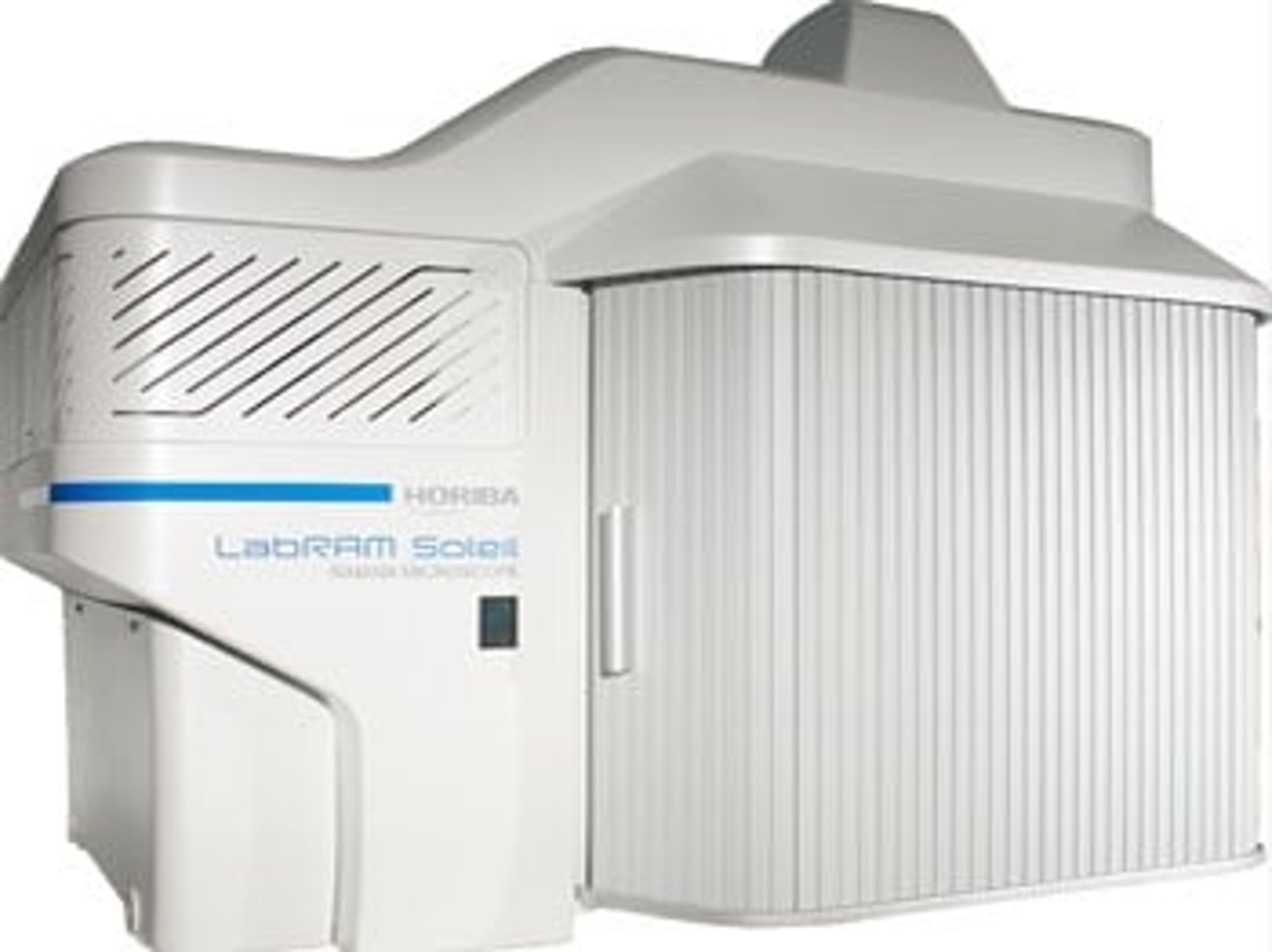
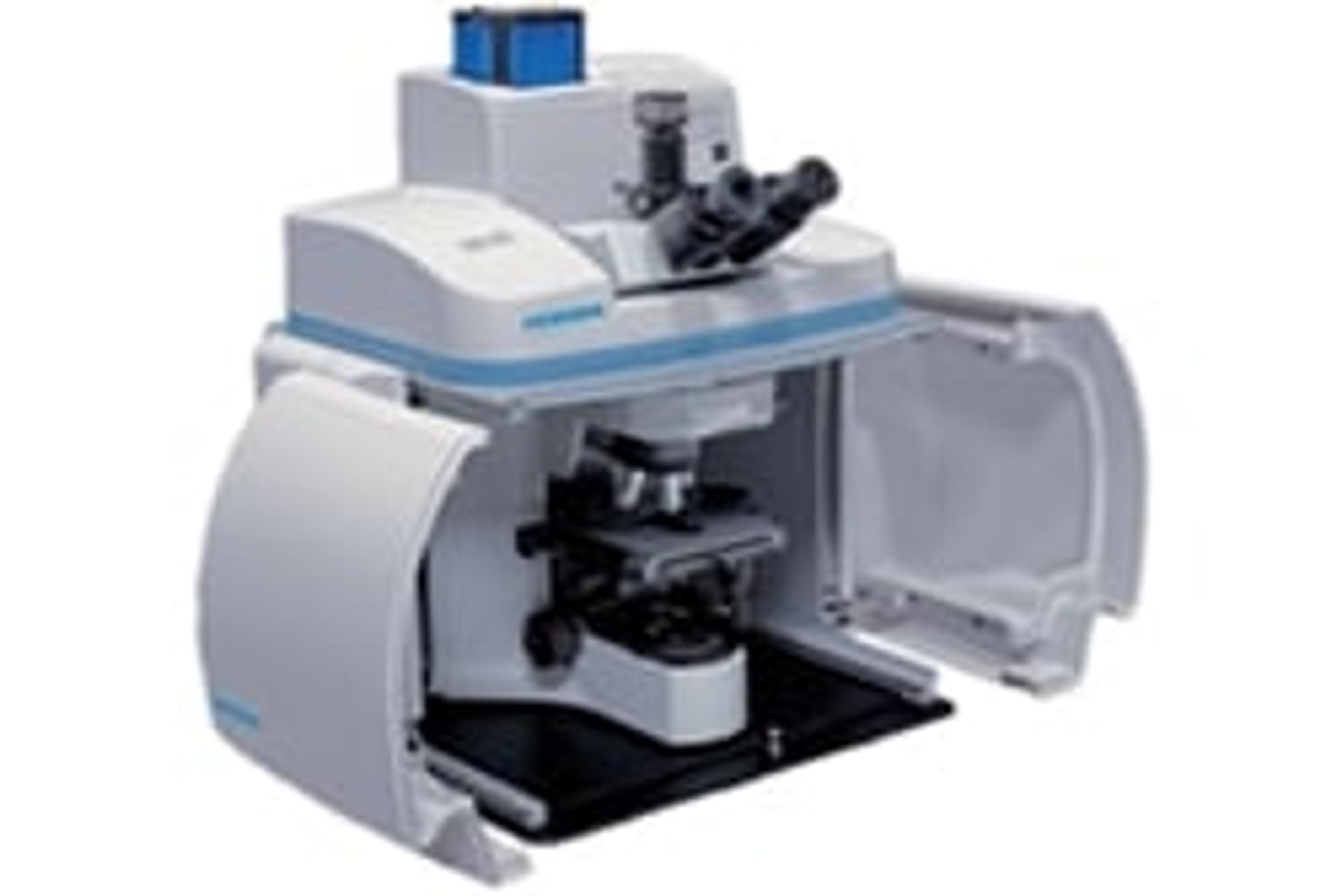
Raman spectroscopy (RS) has evolved into one of the most widely used spectroscopic techniques for material characterization. This non-destructive tool is applied to identify molecules and provide vital information on sample crystallinity, vibrational modes, chemical and crystal structure type, as well as the presence of local stress, defects, and impurities. Characterization is achieved by irradiating a sample with a beam of photons, which interacts with the molecular vibrations and phonons composed within a sample. Typically, this technique is combined with confocal microscopy to provide Raman imaging with micron-scale resolution in either two or three dimensions.
In this on-demand SelectScience webinar, Dr. Adam Holland and Dr. Steven Booth discuss the advances in hardware and software that have made today’s instrumentation accessible to non-Raman experts. Booth also reveals how Raman imaging has helped others obtain a better understanding of products and processes within an industrial setting.
Register now to watch the webinar at a time that suits you or read on to find highlights from the live Q&A session.
Watch on demandQ: Can you use LEDs for Raman?
AH: In principle, there is no reason why not. In practice, LEDs have a relatively wide bandwidth, which means that all the peaks in your Raman spectrum will be shifted, and so broadened by the same amount as the light source. In practical terms, you’ll need a laser for the monochromaticity. So, the answer to that question is not yet.
Q: Could you elaborate more on the selection rule when using infrared (IR)? What are the advantages of Raman microscope versus infrared microscope, particularly when analyzing multi-layered structures?
SB: What I meant by the selection rule in IR is that something is only infrared-active when you have a change in dipole moment during the vibration. Something is inactive when you get a change in polarization. It means the two techniques are quite complementary if you ever use them in tandem. You get different information from both.
In terms of a Raman microscope versus an infrared microscope, the main advantage of the Raman microscope is its specificity. Since you're using white light, you can get down to the diffraction limit of light in that region, which is about a micron. If you are using infrared, you're using longer wavelengths, which is around about 10 microns, so you're automatically sampling a larger area. If you've got thin layers of one or two microns, you're not going to detect those in an infrared microscope.
Q: Can Raman be used to detect COVID-19?
AH: In principle, if there was enough material, you may get a signature from the virus. With our tip-enhanced Raman spectroscopy, you can get down to those sorts of levels. It’s fair to say you would need a lot of research to show that it was COVID-19 and not another closely related viral form. The time that it would take to make the measurement would mean that it's probably not time-effective against something like a rapid PCR test technique.
Q: My samples are dissolved in water. I have tried the related FTIR technique previously but did not get any results. Would Raman be better?
AH: Yes, is the short answer to that. In many cases with FTIR, the signal is dominated by water, which is FTIR-active, but it's only very weakly active in Raman, which means Raman is much more suitable for analysis of aqueous solutions with small amounts of active ingredients in them.
Q: Is there a rule of thumb to define the step distance in a Raman map
AH: Yes. That really comes down to what spatial resolution you want to achieve. In a standard point-by-point map, you would need to have twice the number of data points to start to resolve the smallest of features. It really comes down to, what's the smallest thing you want to measure? The trade-off is always against the time that you're able to spend to make the measurement, which is the reason why we've looked at prioritizing the measurement spots into the areas which are showing rates of change so that you can get the best of both worlds.
Q: What kind of laser wavelength of Raman should be used for polymers such as PMMA, PI analysis?
SB: I think it depends on the polymer and what additives you've got indexed. Generally, you want to minimize the fluorescence background you’re getting with your laser. You will get more fluorescence with the shorter wavelengths, but ultimately that depends on if your polymers will fluoresce. If they will, then you would want to move to a higher wavelength. I'm afraid there's no hard and fast rule of which laser you should pick. It will be dependent on your sample and how much it fluoresces.
Q: Graphene is prepared on the copper plate by the CBD method. Is it possible to analyze the properties of graphene by Raman?
AH: Yes. If you measure it directly on the copper plate with the traditional green lasers at 532nm, there is a bit of fluorescence in that system. What we would recommend is to subtract that out and get the analysis there. Alternatively, if you go into the blue with the bluer laser, the fluorescence is no longer a coincidence with the Raman. You would then get around a lot of those issues, and it's significantly easier to characterize.
SelectScience runs 10+ webinars a month across various scientific topics, discover more of our upcoming webinars>>