Real-time analysis of cellular phagocytosis; Characterization of anti-CD47 antibodies as potential novel immuno-therapies
In this popular on-demand webinar, learn more about novel assays being used to identify new CD47 and phagocyte modulators
25 Sept 2019
Alongside intense efforts to exploit T-cells as immunotherapies for cancer (e.g. checkpoint inhibitors, CAR-T, T cell metabolism), researchers are increasingly considering other immune cell types for novel targets. One example is the enhancement of macrophage function via inhibitors of CD47 “don’t-eat-me” signaling proteins that enable tumor cells to evade clearance by neighboring phagocytes. In order to identify new CD47 and phagocyte modulators, novel, direct and validated assays with throughput commensurate to drug discovery are required.
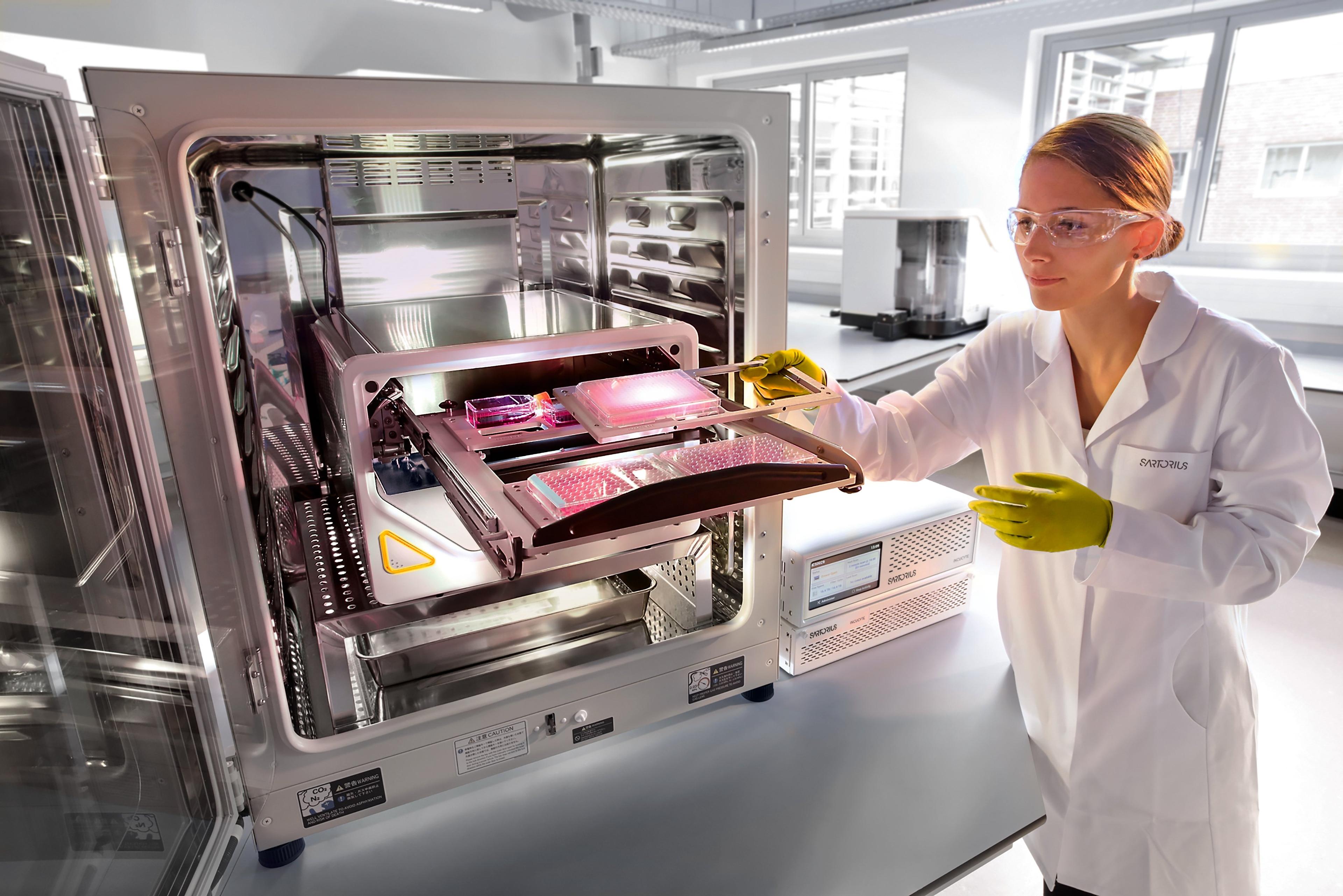
In this SelectScience webinar, now available on demand, Dr. Tim Dale, Head of BioAnalytics Applications at Sartorius, discusses how real-time live-cell analysis using pH-sensitive fluorescent probe-labelled target cells can be used to visualize and quantify phagocytosis and how other live-cell phenotypic assays are used to validate mechanisms of action.
Read on for highlights from the webinar Q&A session or register to watch the webinar on demand.
Watch Webinar Now
Q: How do you induce apoptosis in Jurkat cells? How do you measure that?
TD: Apoptosis is induced by treating with 10 uM camptothecin. This is then removed before addition to the effector cells. Annexin V reagents can be used to demonstrate apoptosis in cells.
Q: Do the labelled target cells cause increased background fluorescence?
TD: Labelling of the target cells can cause a small increase in background fluorescence, but this can be filtered out. The dye is minimally fluorescent in pH 7.4 media and highly fluorescent at pH 4.0 when the cells are engulfed.
Q: Can you count engulfed cells i.e. generate a phagocytic index using this method?
TD: No. The fluorescent signals of engulfed cells are difficult to separate, so we use the total red object area (i.e. the sum of the areas of engulfed cells) instead.
Q: Anti-CD47 is reported to cause externalization of phosphatidylserine (PS). Why is no Annexin V fluorescence observed in the presence of anti-CD47?
TD: Other mechanisms can also stimulate cell phagocytosis, for example increased expression of surface calreticulin.
Q: Have you attempted cellular phagocytosis assays using adherent target cells to mimic solid tumors?
TD: Yes, preliminary data is under development. Initial observations highlight cell size is critical to pHrodo label concentration (or cell density whilst labelling) and more consideration of background fluorescence is required. Additionally, the amount of engulfment is lower than observed for non-adherent target cells, therefore the use of lower magnifications (4x) may be of benefit to increase the number of phagocytic events within the field of view.
Q: Can the IncuCyte phagocytosis assay be used to visualize phagocytic uptake of exosomes by macrophages? If so, can you share a protocol?
TD: Yes, although we do not have a detailed protocol, one could always label exosomes with the IncuCyte Red Cell Labelling Kit. The labelled exosomes would then increase in fluorescence intensity once phagocytosed.
Q: How much time will there be between engulfment of the cell and the breakdown of the pH-sensitive dye by the macrophage, or is that irrelevant?
TD: The pH sensitive fluorophore is not broken down by cells but rather may be recycled out of the cell. This would be cell type dependent. Live-cell analysis enables the time course of engulfment to be monitored, so appropriate timepoints can then be examined further.
Q: Did you block FcR on the macrophages to eliminate the effect of antibody-dependent cellular cytotoxicity (ADCC)?
TD: No - no evidence of ADCC was observed in the data or images.
Q: How do you discriminate with the IncuCyte between trogocytosis and phagocytosis?
TD: By examining the fluorescence signal it would be difficult to discriminate between these. However, by using total confluence or cell count, the engulfment of whole cells can be quantified.
Find out more on this topic by watching the full webinar on demand>>
SelectScience runs 3-4 webinars a month across various scientific topics, discover more of our upcoming webinars>>