Researchers Use Bioluminescent 3D Cell-Based Assays to Mimic In Vivo Cellular Physiology
Find out how scientists at the University of Bologna, Italy, are using Elplasia plates of KURARAY for more predictive drug screening processes
24 Oct 2017

Bioluminescent 2D cell-based assays are well-established drug screening tools. Unfortunately, 2D cell cultures, often, have limited predictive value as they do not reflect the morphology of living organisms. Associate Professor Elisa Michelini, Chemistry Department “G. Ciamician”, University of Bologna, Italy, and her team are using 3D cell models to mimic in vivo cellular physiology. SelectScience® speaks to Prof. Michelini to find out more.
SS: Briefly introduce yourself and your place of work
I am an Associate Professor in Analytical Chemistry working in the Department of Chemistry “G. Ciamician” of the University of Bologna, Italy. I’ve been working at the interface of analytical chemistry and biotechnology for more than 16 years, after first starting to work on bioluminescence during my Ph.D.
The background of our team is highly heterogeneous, taking advantage of different areas of expertise in the development of new bioluminescent tools for biosensing and portable devices (Dr. Luca Cevenini), mammalian cell biosensors (Maria Maddalena Calabretta, Ph.D. student), and yeast biosensors (Antonia Lopreside, Ph.D. student). With the longstanding experience of Prof. Aldo Roda, my mentor and pioneer of bioluminescence and chemiluminescence, we are, now, able to develop cell-based assays and cell biosensors exploiting bioluminescence detection with unprecedented capabilities in terms of analytical performance and multiplexing.
SS: Please describe your work and the need of your current project
Bioluminescence, which is the light emission as a consequence of a chemical reaction occurring in a living organism, is a fascinating natural phenomenon and can be observed in fireflies and marine species. The amazing thing about bioluminescence is that it can be reproduced in the laboratory and exploited to “enlighten” patho-physiological processes, such as intracellular signaling cascades, receptor activation, infections, cancer progression etc., thus providing highly valuable quantitative information.
This can be achieved by exploiting cultured cells, either microbial or mammalian cell lines that can be genetically engineered and reprogrammed to emit light as a consequence of activation of specific molecular targets. Such targets can be receptors for natural hormones, heavy metals, or intracellular pathways whose activation is triggered by inflammatory events, oxidative stress, genotoxic damage etc.
Moreover, it is possible to tune the emission properties, by simply playing with synthetic biology and organic chemistry, and switch the color or change emission kinetics. This provides a formidable arsenal of biosensing tools with exquisite sensitivity and high versatility. These assays can also be implemented into portable miniaturized formats for point-of-need applications (https://www.ncbi.nlm.nih.gov/pubmed/27853830).
We are now moving on from bioluminescent cell-based assays based on conventional two-dimensional (2D) monolayer cell cultures, to 3D cell models. Cells grown in 2D cultures do not often reflect the morphology and functionality of living organisms; therefore, they are not the best model for preclinical screening of drugs. 3D cell models are, thus, required to reproduce a cell microenvironment that mimics in vivo physiological conditions, such as spatial and temporal dynamics, cell-cell communications and physical interactions.
SS: Which Elplasia plates do you use in your research?
We used several formats including 24- and 96-well non-adherent round/square bottom cell culture plates and tested different cell lines, including human hepatocellular carcinoma (HepG2), Hela cells, human epithelial colorectal adenocarcinoma cells (Caco-2), and human embryonic kidney cells (HEK293). We recently tested the feasibility of a bioluminescent 3D assay for inflammation using 24-well round bottom plates and genetically engineered HEK293 cells.
As analytical chemists, we can say that the results (https://www.ncbi.nlm.nih.gov/pubmed/28084029) are highly promising, with the possibility of implementation in any laboratory equipped with basic cell culture facilities. We are, now, testing the multiple pore type plates with different cell lines and preliminary results seem encouraging as well. These plates provide a very straightforward procedure for efficient and reproducible recovery of spheroids for downstream analysis.
SS: What are the advantages of using these plates?
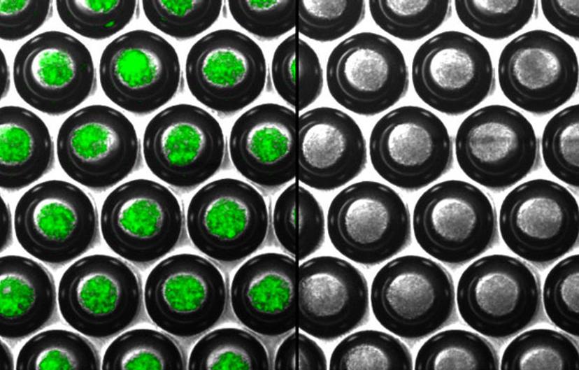
We obtained very homogeneous spheroids using both round and square bottom cell culture plates, with a size suitable to avoid necrosis in the core; and, to maintain the functionality of the cells within the aggregate. We were, also, able to transfect spheroids with good efficiency — that is surely a plus for future development of 3D reporter gene assays.
These plates enabled us to develop a convenient and easy non-lysing imaging procedure for longitudinal monitoring of spheroids. This will pave the way for the development of new 3D assays for drug screening, drug delivery studies etc.
SS: What is the future of your research?
After the first proof-of-concept of bioluminescence imaging of spheroids, there are plenty of applications to explore. It goes without saying that 3D cell models, if developed in a robust and reproducible way, will definitely replace cell-based assays relying on 2D cell cultures. However, the transition from 2D cell-based assays to 3D models is not trivial, and several issues will require rigorous standardization, such as efficient transfection and the implementation of non-invasive longitudinal imaging techniques.
The combination of Elplasia plates and bioluminescence detection provided an excellent basis for obtaining a facile protocol for real-time imaging of live spheroids. Moreover, thanks to the non-lysing nature of the assay, repetitive measurements on the same sample can be performed to monitor protein-protein interactions, cell viability, and cell-cell communication. These assays could be implemented in any laboratory equipped with basic cell culture facilities.
Get more great content like this sent to your inbox: Register as a SelectScience member today