Smaller, smarter, and more affordable live cell imaging systems
In this technical article, Blue-Ray Biotech explores the new innovation of microscope-in-incubator and outlines its key benefits
5 Sept 2019The new microscope-in-incubator innovation, which takes advantage of burgeoning technologies, is designed to provide a more effective and efficient platform for live cell imaging researches at a more affordable price. This promises for more experiments to be conducted within the same budget, so that more scientific questions can be addressed to make the world better in many ways, including the acceleration of the pace of new drug and therapy discoveries.
Live cell imaging is the study of living cells using imaging systems involving microscopy and content analysis, which allows scientists to observe the dynamic changes of cell interactions in real time and over time, in order to provide critical insights into the operations of cells. Driven by new technologies and surging needs for drug discovery, stem cell and other cell biology researches, the global live cell imaging market has quickly heated up in recent years and is expected to reach US$ 7 Billion by 2022, as forecasted by RNCOS in a report released in 2018.
Keeping cells alive and observing cells in real time and over time
Apart from the traditional microscope which observes a snapshot of fixed cells without the ability to look at dynamic biological processes, a live cell imaging system adds an incubator to the microscopy system to keep cells alive and allows for long-term experiments on the dynamic processes of how cells translocate, interact and respond to environmental cues.
This incubator mimics the physiological conditions optimal for the cells’ health so as to ensure the cells not only stay alive but remain in a metabolic state with no nonspecific changes that could possibly alter the process being observed. For example, human cells need to be cultured under an environment near and above 37℃ with 5% CO2 concentration as if in a human body, along with other conditions.
Therefore, a live cell imaging system must contain basic components that fulfil the following requirements:
Incubator that keeps cells alive throughout the observation process
Microscope for observing the cells
Imaging device that records cell images over the process
Analysis software that converts quantitative image-based data into significant insights
Incubator-on-microscope, an option of luxury
There are currently two methods for a microscope to incorporate cell culture device. One builds a specialized incubation and imaging chamber onto a general-purpose inverted microscope. This method preserves complete functions of a traditional microscope and allows for multiple illumination and camera options and also higher image quality. However, considering on the physical constraints of the microscope, the stage-top incubator has to be reduced to a smaller size than a normal cell culture chamber, and it is more difficult to control environmental variables within a smaller space, entailing environmental stability and cell viability issues.
Most important of all, a traditional full function microscope with an add-on specialized incubator could be as pricy as US$50,000 to $100,000 per unit, and a whole unit could be occupied for days, weeks or months just for one single live-cell experiment, making it economically irrational.
Microscope-in-incubator, a dedicated system improving convenience
The other method for microscopy integrating incubation is a newer innovation in the market, which builds one or several inverted microscopes with reduced functions merely for live-cell imaging and monitoring purposes, and installs the dedicated system in any lab’s existing cell culture chamber, with a control unit connecting to the specialized microscope and networking system.
Thus, users may adjust lens focus remotely without opening the incubator and creating a disturbance of cells, and observe cells in situ in a non-perturbing environment without the need to remove them to instruments and compromise cell health. Meanwhile, all images will be automatically and continually recorded day and night, and researchers may view these images anytime without missing any details.
With simplified functions and image quality level acceptable for live cell researches, such dedicated microscopes are usually provided with lower expandability and on lower prices, suited for experiments relying on long-term live cell imaging. Besides, users do not have to purchase an additional incubator for live cell imaging researches, so the overall cost for live cell imaging can be reduced to a more affordable level, giving economic benefits to labs and researchers.
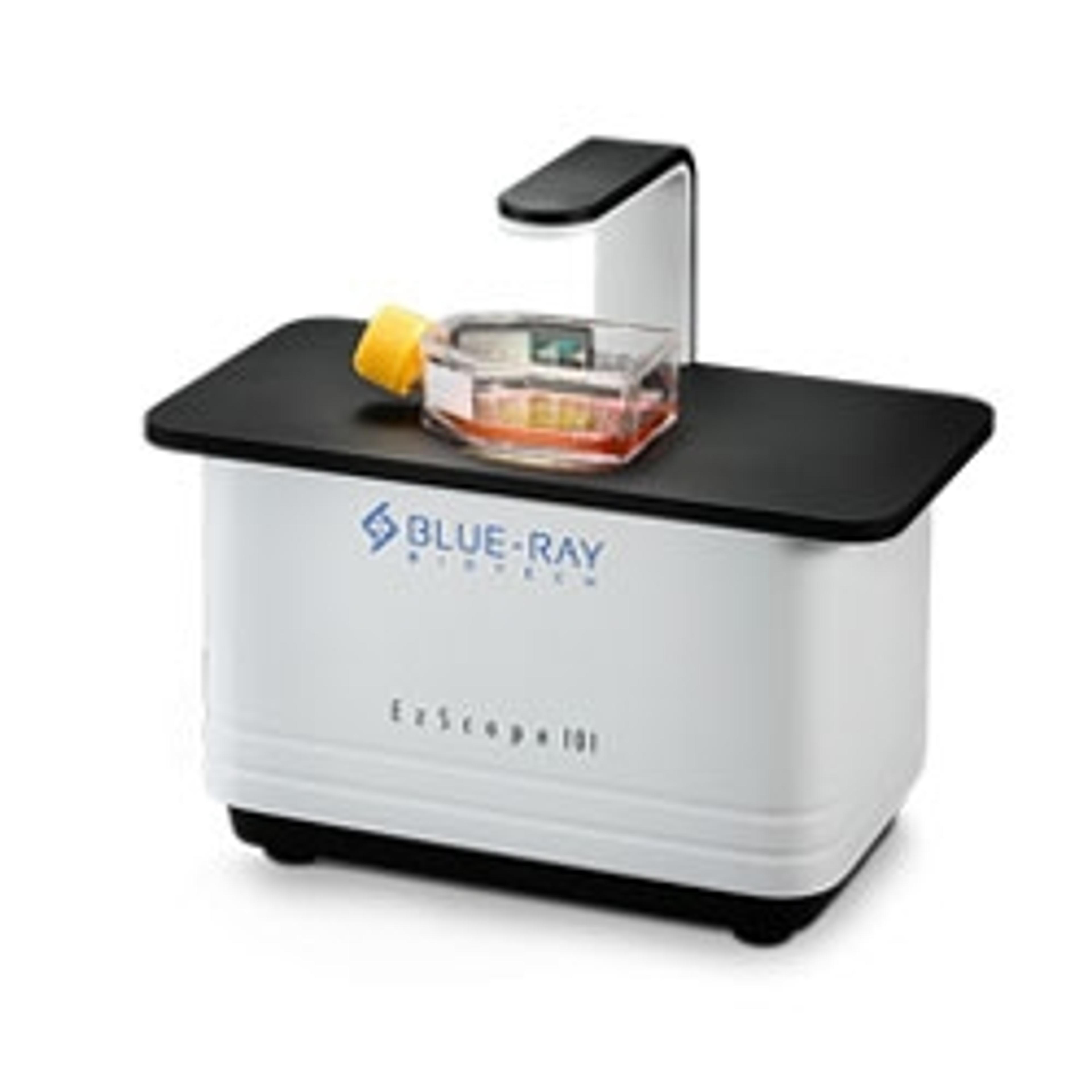
EzScope 101streamlines research workflow from incubation to presentation
The Blue-Ray Biotech EzScope 101 Live Cell Imaging System is one of the products best exemplifying the up-to-date innovations of the next generation live cell imaging technology. Featuring a compact size with reinforced construction, the EzScope 101 is highly compatible and can be fit into most incubators used in labs. It allows for connection of up to 4 units of devices in the same incubator, allowing for simultaneous multiple cell experiments with precisely controlled conditions.
Though some microscope-in-incubator imaging system allows for rather low expandability, the EzScope 101 still manages to improve flexibility by providing two exchangeable lenses. In addition, the EzScope 101 is one of the rare products in the market which provides the feature of motorized focusing, allowing users to remotely adjust lens focus via a software-enabled dashboard installed on the computer without the need to open the incubator for focusing, thus avoiding disturbance of cells.
The EzCapture software provided by EzScope, not only allows users to build a dashboard for viewing and editing cells images, but also provides functions for analyzing results acquired by the device. With a single click on function menu, users may easily implement cell coverage estimation, cell growth rate determination and cell migration measurement, which are often applied in assays for wound healing, stem cell behaviors and intravital studies, and turn the data into presentation-ready images and videos. Researchers may view the cell images in real time or later, enjoying walk-away convenience and improved productivity.
New products accelerating new discoveries in the cell world
Leveraging new technologies, live cell imaging system now has become smaller, smarter and much less expensive, which will make it more convenient and affordable for scientists to conduct experiments either in basic or applied researches for a wide range of purposes, from oncology, immunology to new drug and therapy discoveries, helping to accelerate progress in these areas and improve human life. For those labs frequently and intensively occupied in live-cell imaging assays, cell-imaging systems that employ dedicated microscopes installed in incubator are obviously a more ideal option over expensive stage-top models.
For more science news, straight to your inbox, join SelectScience today >>