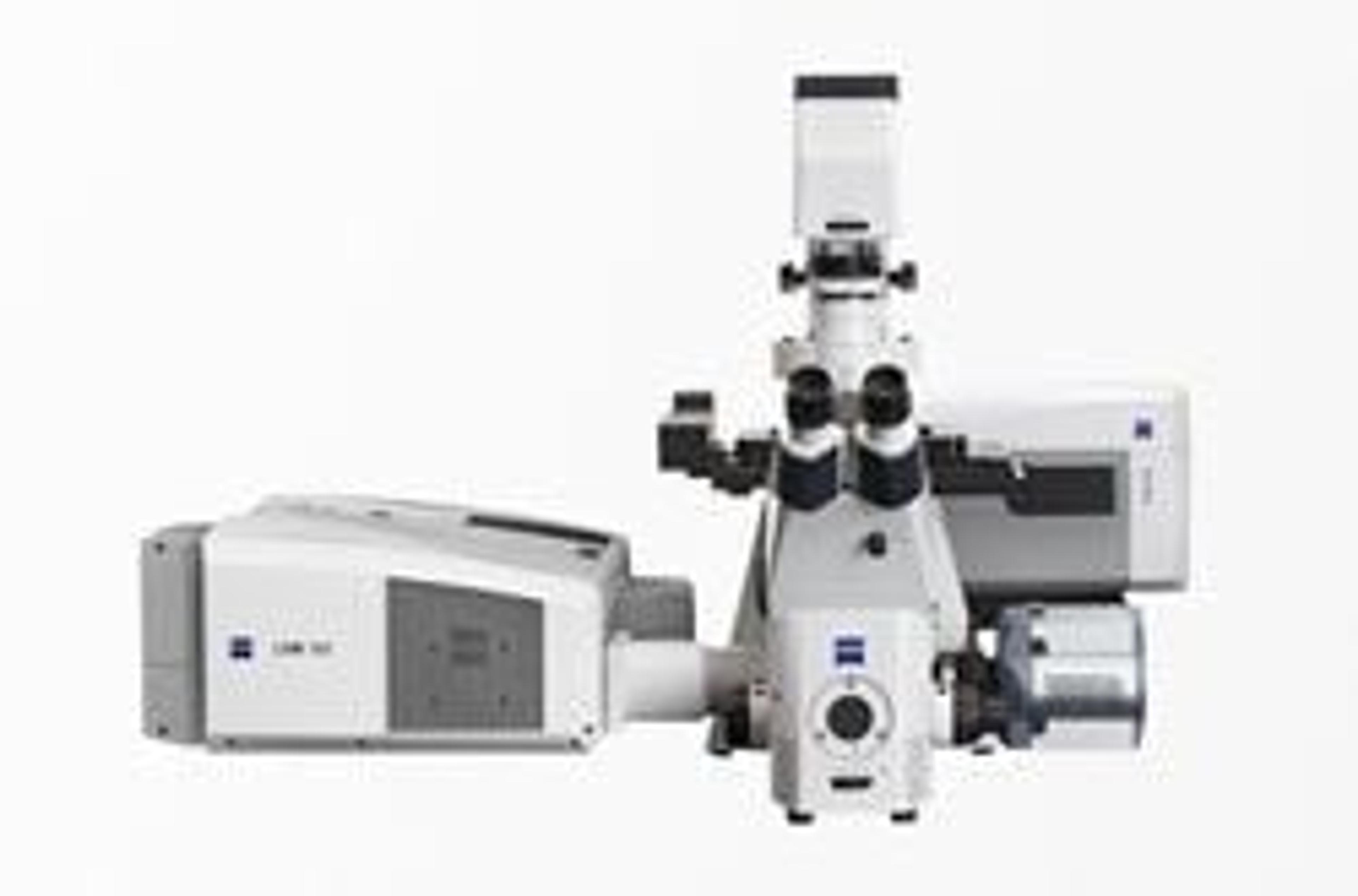
Superresolution with Microscope Systems from Carl Zeiss
16 Oct 2009Carl Zeiss announces the ELYRA product family that allows the superresolution PAL-M and SR-SIM microscopy techniques to be comprehensively used and individually combined for the very first time. This enables scientists in biomedical research to image cellular structures marked with fluorescent dyes with maximum spatial resolution below the classic diffraction limit of microscopes.
The system line comprises three microscope systems: ELYRA S.1 (SR-SIM) is the first microscope system to allow the use of SR-SIM technology on the basis of a standard microscope stand. With ELYRA P.1, PAL-M technology is now being offered commercially for the first time. The ELYRA PS.1 combined system permits the very first combination of the two technologies on the same stand, both with each other and with a laser scanning microscope. This means that an object can be imaged with the three LSM, SR-SIM and PAL-M techniques in succession.
ELYRA gives users the opportunity to acquire the superresolution systems on either a “turnkey” or modular basis. They no longer need to invest in single extremely specialized and expensive systems. PAL-M technology offers the highest resolution currently available while the outstanding feature of SR-SIM technology is its high level of flexibility in the choice of dye. This means that both superresolution methods are available without the previous limitations in dye selection or user-friendliness.
These outstanding properties have been made possible by the new laser widefield illumination with fully motorized elements for illumination in Total Internal Reflexion (TIRF) and for the superresolution structured illumination (SR-SIM). Electron multiplying CCDs guarantee highly sensitive detection. Intuitive operation and comprehensive data analysis are implemented with the ZEN software.
The optimal combination for high-end research is offered by ELYRA in conjunction with the Laser Scanning Microscope 780. However, PAL-M and SR-SIM can also be combined with the LSM 710 laser scanning microscope.

