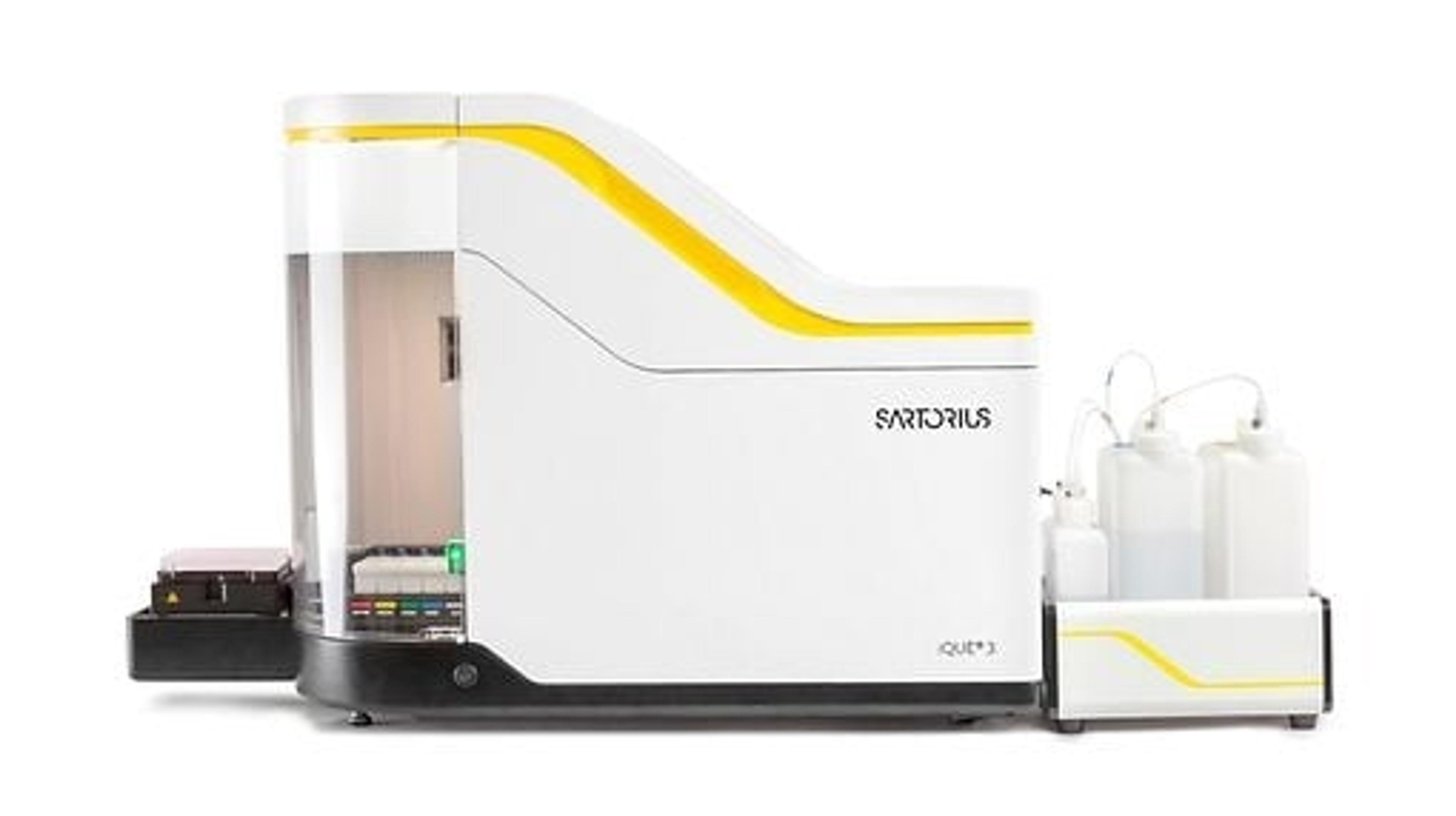
T cell characterization combining advanced flow cytometry with live-cell analysis
Watch this on-demand webinar to gain insight on how advanced flow cytometry and live-cell analysis can facilitate faster drug development
24 Aug 2020

Harnessing the power of the immune system using therapeutic drugs has enabled a huge advancement in the fight against cancer. T cells are the focus of this immuno-oncology field, as they can be selectively activated by therapies, causing them to seek out and destroy diseased cells.
In vitro models can enable a deeper understanding of T cell biology. Knowledge of the intricacies of this immune sub-population function may be used to facilitate predictions of the effectiveness of therapeutics for patients. Therefore, in vitro models can promote more efficient and faster drug development.
In a recent SelectScience webinar now available on demand, Dr. Clare Szybut, Senior Scientist, Sartorius Group, provides insight on the following topics:
- How advanced flow cytometry can be used to link T cell function to marker expression and cytokine release
- The characterization of bispecific antibodies to reveal mechanistic insights to actions on T cell activation, killing, exhaustion, and memory sub-populations
- How live-cell analysis can complement advanced flow cytometry to provide additional insights into T cell function
Read on for highlights from the Q&A discussion at the end of the live webinar or register to watch the full webinar on demand.
Watch on demandQ: Regarding activated T cells, we know that some donors/patients do not respond to aCD3. Have you tested the combination aCD28/SEB versus aCD3/aCD28 versus SEB?
CS: No, we have not tested this combination, we have however tried activating with stem cell technology ImmunoCult CD3/CD28/CD2 which gives very good activation and provides a good alternative to Dynabeads.
Q: Have you also developed and validated mouse Kits?
CS: Yes, we have a mouse kit for the T cell activation profiling kit which has been validated.
Q: Can adherent target cells be used with these assays?
CS: Yes, we have successfully tested a number of adherent cell lines. The incubation with Accutase described in the combined workflow allows for adherent cells to be lifted so they can be processed by the Intellicyt® iQue®.
Q: Can chemokines be monitored as well as cytokines?
CS: Yes, chemokine release can also be monitored and added as an auxiliary agent in the same way as extra cytokines can be assayed for. Our website lists all of the chemokines that can be detected.
Q: You have developed kits with pre-gating strategy. Are those kits compatible with other flow cytometers?
CS: These kits are best suited for use on the iQue® platform as the compensation and FMOs have already been performed. As we do not specify the identify of antibodies we use or concentrations it would be difficult to translate over to another flow cytometer.
Q: Is the iQue® kit only useful with well plates? Can it be utilized using flow tubes as well?
CS: Yes, it can be used for tubes as well, however we recommend plates as this increases throughput. If using tubes, it needs to be 1.5 ml Eppendorf tubes, not conventional flow tubes.
Q: Can other cell types be used to express CD19 for the BiTE interaction?
CS: Yes, we have also used Raji cells. In theory any cell that is positive for CD19 should allow the BiTE to bind and illicit an immune response.
Q: What is the ratio of the beads / cells you tested?
CS: We used a top concentration of beads to cells of 1:1. For exhaustion we recommend 3:1.
Q: What types of immune cell preparations can be assayed?
CS: These assays are capable of identifying T cell populations from isolated PBMCs, but T cell isolations can also be used as well as CAR-T preparations. These kits are designed specifically to characterize T cell subpopulations however we also provide kits for monitoring other subpopulations such as natural killer cells.
Q: Can temporal data be collected using the kits as well as single end points?
CS: Yes, multiple time points can be collected so that time course data can be achieved. It is sometimes recommended to do this over the course of stimulations so that the temporal differences in when markers start to be expressed can differ over time.
Q: Would the activation beads interfere the QBeads detection?
CS: No, the beads do not interfere with the QBeads, they are a different size and the QBeads are detected in the BL2 channel.
SelectScience runs more than 10 webinars a month across various scientific topics, discover more of our upcoming webinars>>

