The Latest Videos for Neuroscience Research
Discover the latest innovations from Neuroscience 2015
20 Nov 2015
Discover the latest innovations from Neuroscience 2015
SelectScience® attended Neuroscience 2015, in Chicago, USA, to bring you exclusive interviews and videos about the very latest technological innovations in neuroscience. Discover how virtual reality technology can help us to investigate neural systems, and how to use functionalized nanoparticles for live tissue imaging.
1. Discover the New EVOS FL Auto Imaging System
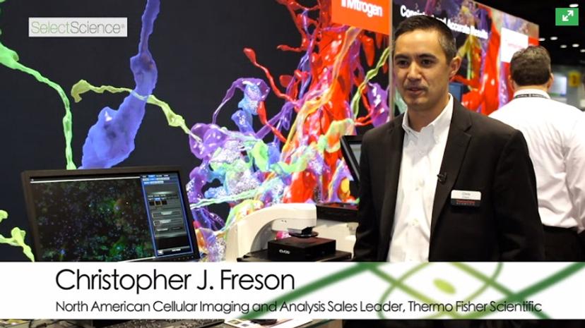
In this video, Christopher J. Freson, North American Cellular Imaging and Analysis Sales Leader for Thermo Fisher Scientific, introduces the new Invitrogen™ EVOS™ FL Auto Imaging System. By avoiding complicated eye pieces that are difficult to operate and instead designing the EVOS FL Auto to work with a touchscreen, Thermo has maintained the image quality whilst making the system easier to operate. Watch video.
2. Meet the Brand New ChemiDoc-It from UVP
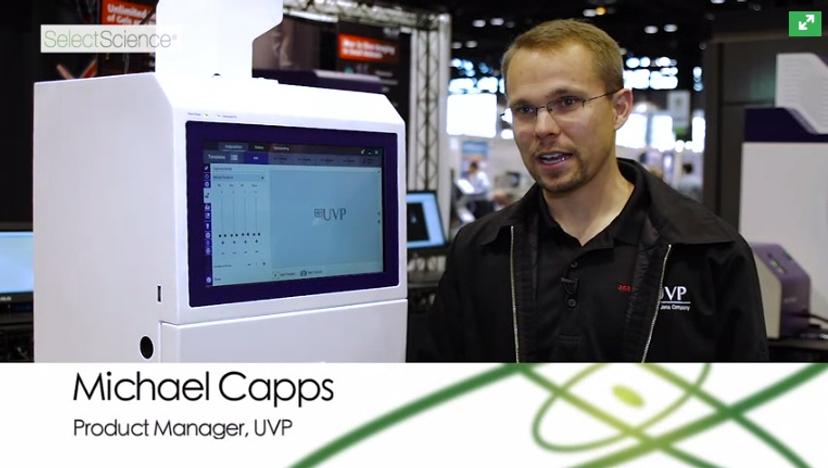
Discover the brand new ChemiDoc-ItTS3, an all-in-one system for imaging chemiluminescent western blots, as well as gels. In this video, Michael Capps, Product Manager at UVP discusses the new features of the ChemiDoc-ItTS3 including a brand new camera and lens, now capable of cooling up to -57°C from ambient, providing superior sensitivity for low light imaging applications. For imaging of red, green, blue, or NIR blots, the modular component BioLite, a spectral light source, can also be added. Watch video.
3. Virtual Reality Visualization of Complex Data Sets Acquired on ZEISS Microscopes
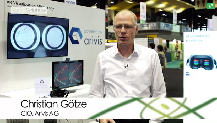
In this video, Christian Götze, Head of Development at arivis AG, explains a new approach to look at the large data acquired with microscopes. ZEISS and arivis partnered to create a prototype which allows you to render and visualize terabytes of volume data with the Oculus Rift virtual reality headset. Gain a deeper insight into very complex brain structures and intuitively understand your data. Step into your sample, fly through a brain, past brain cells and analyze the image from every angle, and from inside. Watch video.
4. Solving the Problem of Neural Population Coding at Manchester University
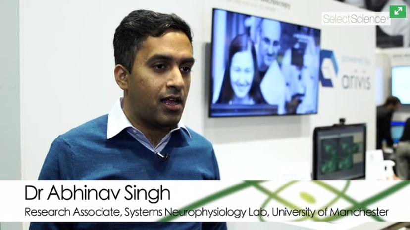
Watch this video to hear Dr Abhinav Singh, Research Associate in Systems Neurophysiology Lab at University of Manchester explain the problem of population coding. When information about the external world is received via sensory signaling, it is transformed by our brain, but exactly how this transformation happens is unknown. Dr Singh is looking into how spikes in the pre-frontal cortex encode information about rule learning, and also discusses the fantastic viewpoint virtual reality offers in data analysis. Watch video.
5. How Large Scale Electron Microscopy is Revealing Animal Behavior at Harvard University
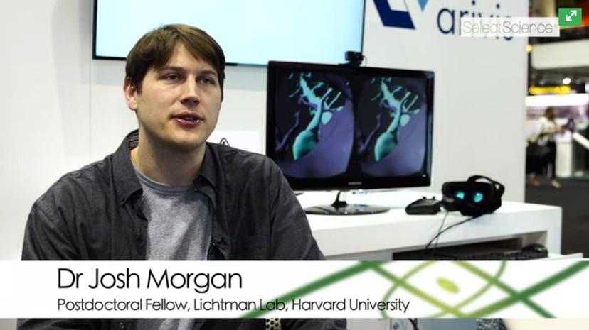
Discover how Dr Josh Morgan, Postdoctoral Fellow in the Lichtman Lab at Harvard University, is trying to understand how the nervous system works by looking at how neurons organize their synapses with one another. All behavior in animals depends on cells talking to each other via specific connections, sending out long processes and forming synapses with each other. Understanding the pattern of those connections is one way of understanding animal behavior. Watch video.
6. Functionalized Nanoparticles to Detect Biological Systems at the University of Berkeley
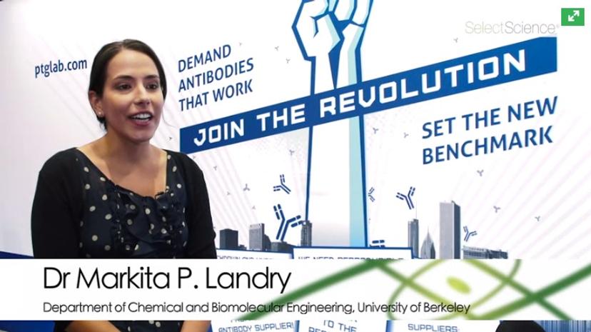
Learn more about how optically active nanomaterials and particles are being used to detect molecular systems in living tissue. In this video, Proteintech travel grant winner, Dr Markita P. Landry, Department of Chemical and Biomolecular Engineering at University of Berkeley, explains how she developed probes that are responsive to specific proteins, emitting NIR when they are near and allowing very dense tissue to be imaged. Watch video.
7. Live Neural Imaging Reveals Neural Interactions Over Time at Cornell University
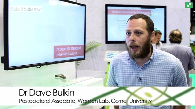
In this video, Dr Dave Bulkin, Postdoctoral Associate in the Warden Lab at Cornell University, explains his research into how neurons work together. This involves imaging cells in the lateral habenula, which is involved in reward pathways, using mice implanted with gradient refractive index lenses from Inscopix. By imaging hundreds of neurons at once, Dr Bulkin is hoping to move on from thinking about single neurons, and instead understand the mechanics of large populations. Watch video.
Discover the latest news, methods and reviews on neuroscience by visiting our dedicated page.