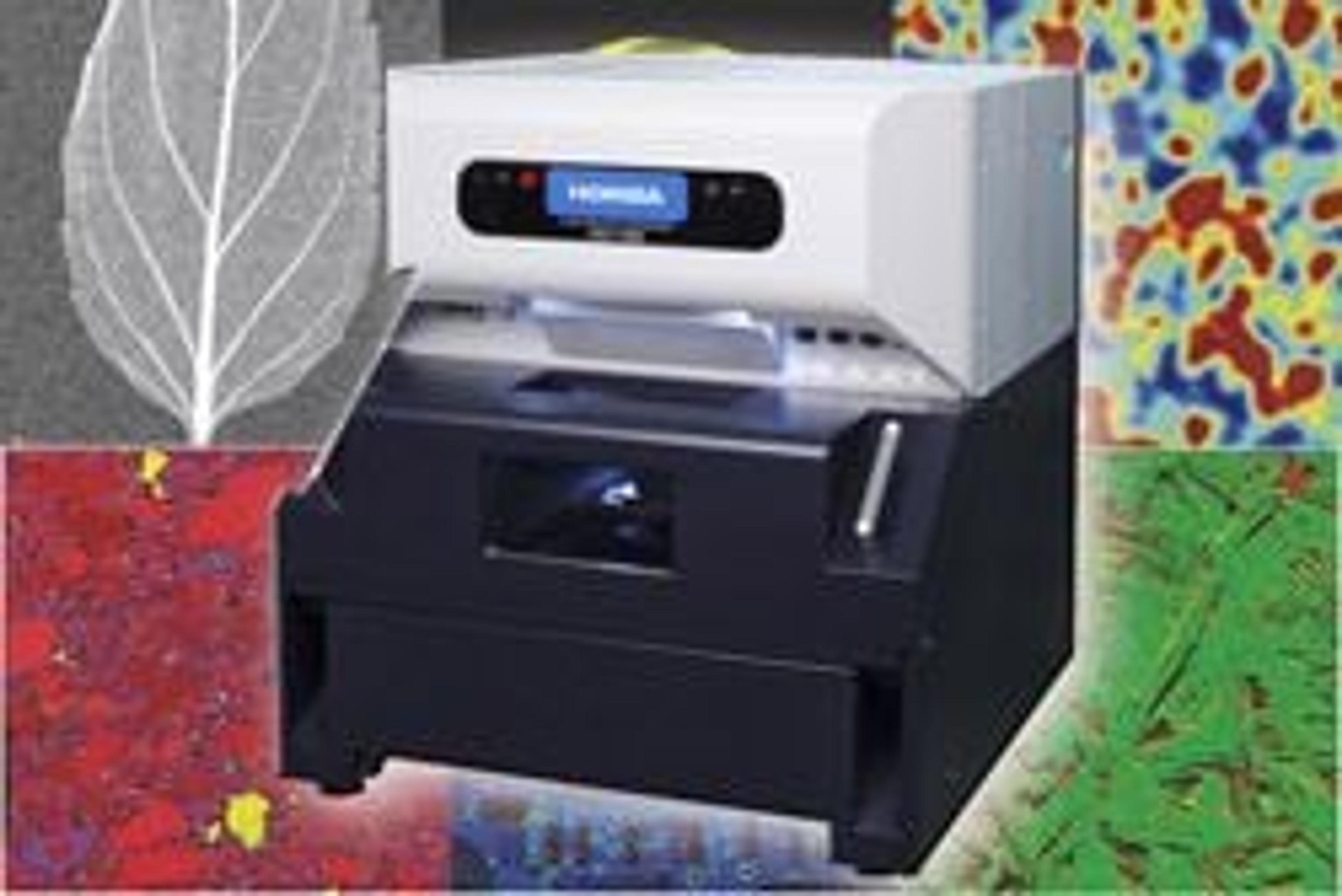
The XGT-7000 XRF Imaging Microscope with 10 μm resolution
9 Sept 2007HORIBA Jobin Yvon is pleased to offer the XGT-7000, the latest system in the XGT series of energy dispersive x-ray fluorescence (EDXRF) microscopes. This ground breaking instrument offers fast, high sensitivity qualitative and quantitative elemental analysis and imaging, transmission x-ray imaging, unique dual vacuum modes, and dedicated imaging acquisition/analysis software.
A wide range of research benefits from non-destructive analysis of discrete microscopic particles and high spatial resolution element imaging. The XGT-7000 provides the answer for applications as varied as forensic science, pharmaceutics, materials, engine wear analysis, geology, museums, archaeology, electronics and the life sciences. A choice of two x-ray beam diameters is available on the system, ranging from 1.2 mm through to a world leading 10 µm. Beams can be switched in a matter of seconds, ensuring complete flexibility on a single bench top instrument.
The instrument’s unique dual vacuum modes offer the researcher complete control for varied analyses. The Full Vacuum mode provides the highest sensitivity for light elements such as sodium, magnesium and aluminium. However, for water containing samples such as biological cells and fragile archaeological objects which can be damaged by vacuum conditions the Localised Vacuum mode allows the full elemental range from sodium to uranium to be analysed with the sample chamber maintained at normal atmospheric pressure.
The XGT-7000 SmartMap software leads the user through project set up, experiment configuration, acquisition and data analysis, and modules such as auto peak labelling, spectral searching, FPM and calibrated quantification and report generation are easily accessed. Elemental images can be generated post-acquisition and adjusted at will, ensuring that the potential of the information rich element image data can be fully utilized.