Webinar Highlights: Increased Confidence of Wound Healing Assays
Find out how to boost confidence in results of cell migration and wound healing studies
21 Nov 2016
Learn how multimode readers are being used to automate wound healing assays

Dr Katrin Flatscher
Application Scientist, Tecan

Dr Michael Fejtl
Market Manager, Detection, Tecan
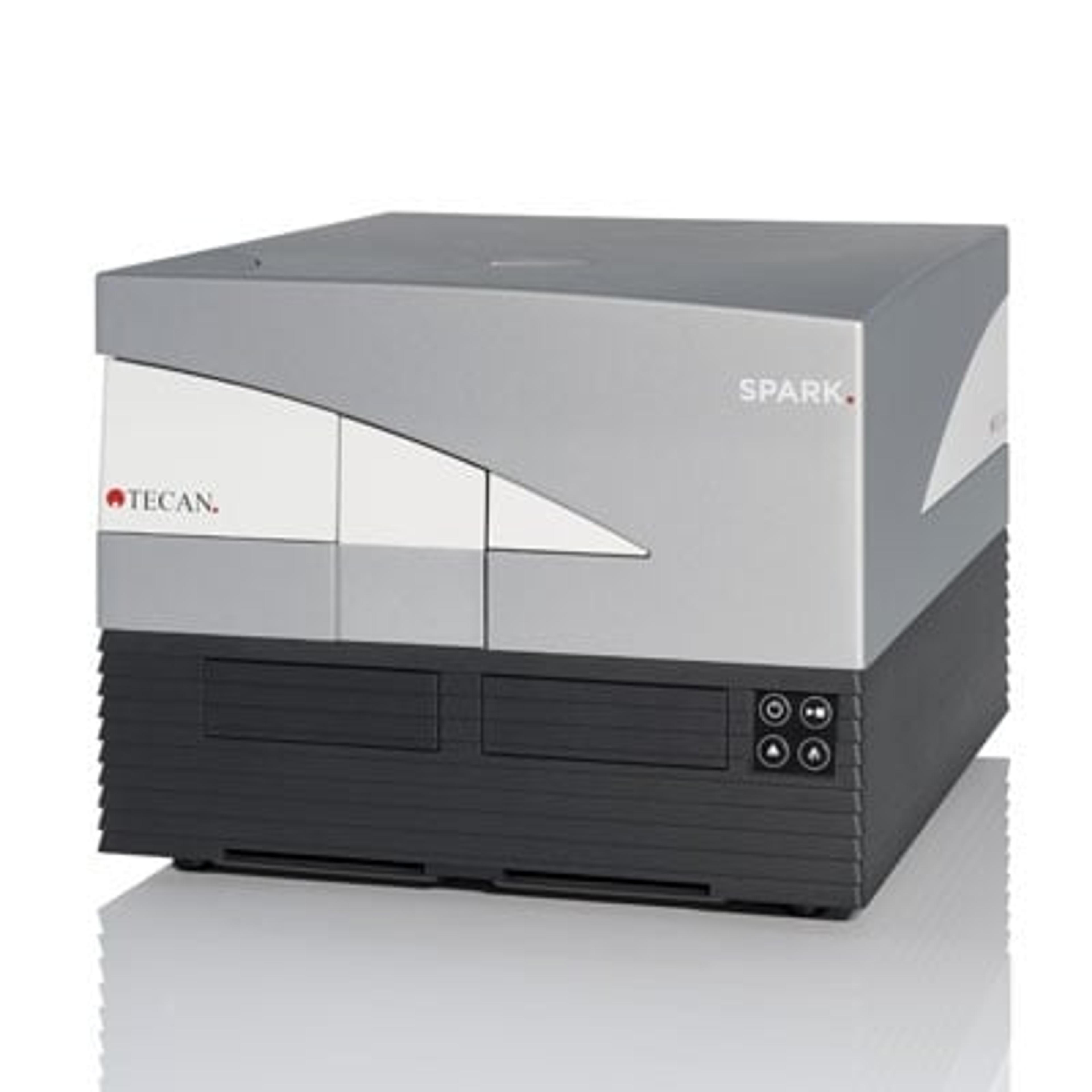
Dr Katrin Flatscher, an Applications Scientist at Tecan, and Dr Michael Fejtl, Market Manager at Tecan, gave an informative webinar on how wound healing assays can be improved through the assessment of cell migration and cell viability in parallel. In the webinar, they explain how the Spark™ 10M Multimode Microplate Reader can be used to measure these parameters throughout the assay and lead to improve the confidence of the results obtained.
Read on for highlights of the Q and A session with Dr Katrin Flatscher, and if you missed it, watch the webinar on-demand.
Q: What advantages does the Spark™ have in comparison to manual cell counting?
A: The biggest advantage of the Spark™ in comparison to manual counting is the increased throughput. The slide adapter allows you to analyze eight independent cells samples in one go. Imagine how long it would take to count eight cells manually under the microscope. Also, it is standard practice to count at least 100 cells manually, whereas the Spark™ is accurate at much lower cell numbers.
Additionally, with the Spark™, no stain, label or dye needs to be added if you simply want to count the cells. Cell viability can also be easily assessed by adding a simple trypan blue stain to discriminate live and dead cells.
Automated cell counting will give your results a much better reproducibility and a standardize you cell based workflows. It allows for a much higher throughput and will save you time in the laboratory.
In a case where the cells have a high speed of migration and gaps in-between the cells, how do you measure wound healing using the Spark™?
A: The Spark™ instrument measures the various indicators of wound healing by taking pictures of the cells on the surface of the microplate wells over the course of the experiment. For cells with a fast migration speed, we recommend taking pictures at shorter intervals. The kinetic measurement function allows the operator to freely set the intervals between pictures, and theoretically, depending on how many wells you are using, you could take one picture per minute.
Gaps in-between will appear in the images taken by the Spark™. The software can discriminate between cells and the well bottom and will give you the confluence value that reflects the percentage of cell-covered area on the selected surface spot.
Q: Can a fluorescent label also be imaged using the Spark™?
A: Currently, fluorescence imaging is not possible with the Spark™ as it has a bright field imaging system which enables analysis of cell number, viability and confluence. However, since the Spark™ is a multimode reader, you can measure fluorescence in parallel with cell imaging. To learn more about using fluorescence, read our application note about how confluence was measured in parallel with cellular GFP signals in a cancer cell model system.
Q: Could the Spark™ be used with the classical wound healing assay?
A: The Spark is compatible with brightfield imaging-based cell migration assays that are normally analyzed using a microscope. Traditional scratch assays can be done with the Spark™, however it is recommended to use the whole-well imaging pattern to analyze the entire scratch area, even if it is not located in the middle of the well.
Q: How would you distinguish cell migration from cell proliferation during the wound healing assay?
A: Wound healing experiments are mostly long-term assays, so there is the potential some degree of cell division throughout the analysis period. There are two solutions to this. The first is using a fluorescent tag or label to distinguish daughter cells. The second method, and that which was used in the experiments described in our webinar, is to prevent cells from proliferating by removing the serum (FCS) from the growth media during the gap closure assay.
Q: How could I determine the cell number via the confluence number?
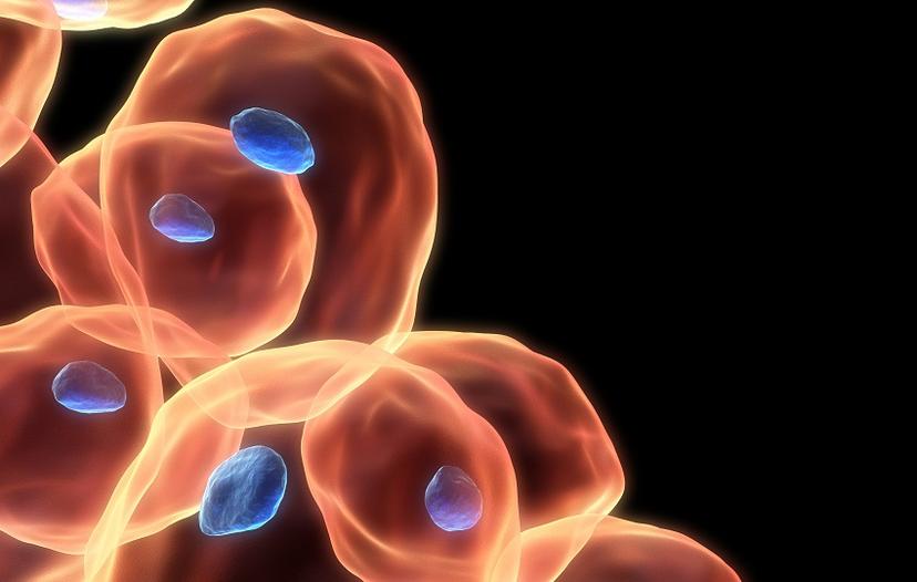
A: Technically, confluence is not the same thing as cell number. Different cell lines can have different cell size and therefore will reach confluence on the given growth area at different cell number. For example, hu-VEX cells have an approximate diameter of 12-15 µm, whereas HeLa cells are closer 20 µm, so a HeLa culture would reach 100% confluence at almost half the number of cells compare to hu-VEX.
To determine approximate cell number from confluence you’d have to create a standard curve which correlates the confluence and cell number at the time of seeding. You can then use this standard curve as a means of estimating cell number from confluence in your experimental assays. The standard curve would be determined by seeding the cells into microplate well, letting them settle and adhere and measuring the confluence. You would then carry out a cell count and note down what number of cells gives that percentage confluence. This would be repeated over a range of confluence percentages and it would also be advisable to replicate the wells and take an average to give better reliability. A separate curve must be created for each induvial cell type due to difference in cell size.