Webinar Highlights: Latest Innovations in Live Cell 3D Imaging with Confocal Microscopy
25 Aug 2016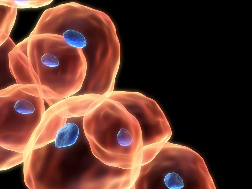
Image - Shutterstock/Sebastian Kaulitzki

Dr. James Lopez, Manager at Olympus Corporation of America
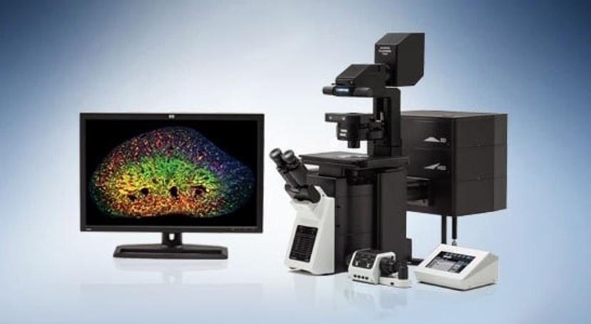
FV3000 Confocal Laser Scanning Microscope The FV3000 is optimized for macro to micro imaging of cells, tissues and small organisms.
Dr. James Lopez is the manager of the Life Science Applications Group at Olympus Corporation, and has a background in Biomedical Research. He presented a webinar with SelectScience® to discuss the issues and challenges associated with live cell 3D imaging, the improvements that have been made with laser scanning confocal microscopy and how the technology is still being developed. Read on for the summary of the Q&A session of the webinar, and if you missed it, watch the free, on-demand webinar here.
Q: I often have problems imaging red fluorescent proteins – the signal is dim or very photo bleachable – do you have any recommendations for this?
A: In my experience, monomeric dyes, like mCherry, are more biologically capable of not interfering with cell behavior. They’re often chosen because they’re smaller than, for example, a tandem dimer with tdTomato. Because you have two molecules of tomato, tdTomato however is brighter and has a little less photo bleaching than mCherry. There are improvements in the red proteins all the time.
The other note on this is that if the system is very sensitive and is optimized for these red proteins, photo bleaching becomes less of an issue. So there is choosing the right dye, looking at the brightness and the photo stability of that protein you’re selecting, and making sure that the system is optimized for those red channels. Some detectors are more sensitive in the green wavelengths and drop off in the red, other detectors are brighter in the red. It’s important that all of these pieces come together for that red fluorescent protein.
Q: How many distinct dyes do you think you can use with spectral imaging?
A: I can speak about the Olympus FV3000. Our algorithm actually has a limit of 16 different colors that it can isolate out. The limitation is the number of detectors and the algorithms used to separate out the number of proteins.
In some transgenic fish expressing Brainbow, or Confetti, there can be many, many hundreds of different combinations of chromophores. As far as I know no system is capable of specifically discerning that many different hues or variants of color. But certainly 16 different fluorescent proteins are possible on a system like the FV3000, and 16 is really a high number. There may be some systems on the market that are not able to do 16 and there may be some that can do slightly more, but usually right around 16 is the theoretical limit as a practical every day experience.
However, when people put in more than 4 or 5 colors into a sample usually it’s a struggle; it may be possible with a lot of work, but usually 4 to 5 different proteins or dyes in a different sample simultaneously is the practical upper limit of what people are actually doing successfully.
Q: How would you need to prepare plant samples, since plant tissue is thicker in comparison to other samples?
A: Plant tissue is very difficult, as plant tissue has a lot of auto fluorescence and many people use Green Fluorescent Protein (GFP). GFP and auto-fluorescence tend to occupy the same spectral range, so a spectral system has to be well equipped to be able to separate the auto fluorescence from the GFP. It is worth noting that CFP is in a very quiet spectral region in plant tissue, with less auto-fluorescence.
There are some clearing methods available for plant biology that I was not aware were being used until I started imaging with some of them. There are chemicals and protocols optimized for plant tissue to remove that auto-fluorescence, so you can image deeper into plant tissue with less background. These techniques are still developing and many, if not all of these clearing techniques, are not compatible with live cell biology.
Q: Many confocal systems are available in upright or inverted configurations, have you got any recommendations as to why one system would be preferred over the other?
A: I think that part of the answer really comes back to what is the sample. If you’re imaging a clear specimen that is very, very large and thick, in some situations an upright system lends itself to that application a little bit more.
Electrophysiology in some situations is a little bit easier on an upright. But in general an inverted microscope is more flexible; it’s easier for sample mounting, it’s easier for the researcher, it’s easier for perfusion, and it’s easier to mount a lot of different apparatuses on the stage and then do your imaging that way.
It’s not always a clear cut decision, upright versus inverted – the vast majority of people imaging on a confocal are using inverted systems – but that discussion really comes back to the experiment, the application, and what is the best configuration for that experiment.
Q: How far away are we from a microscopy technique that allows fast super resolution in thick live tissues, and what advances do you think will come in the next five to ten years?
A: FLUOVIEW (FV) OSR technology is capable of imaging into thicker specimens with super resolution but it is not capable of live cell imaging due to the speed.
Most super resolution is still happening very close to the surface and super resolution systems are still struggling to generate frame rates that are equivalent to a non-super resolution imaging speed. Olympus has developed its SD-OSR (Spinning Disk-OSR), which is suitable for both live specimens and relatively deep SR imaging at 100nm resolution or so (depending on the sample).
But the technology is advancing. I think the technology is getting more sensitive, and getting faster. Those two factors are important but many people also, above all, want the system to be easy.
So the Olympus SR technology is being married to ease-of-use which improves user experience, and I think automation has certainly improved throughout the last decade. The automation, the ease-of-use, and the simplification of these systems is probably a very attractive area for continued development as well as the improvements in speed, resolution, and sensitivity.
I think that in the next ten years we will see an increasing ease of use of these systems where you don’t have to be an expert using the system 25 times a year or more to generate beautiful images.