Western Blotting: 10 Technical Tips for Success
8 Sept 2014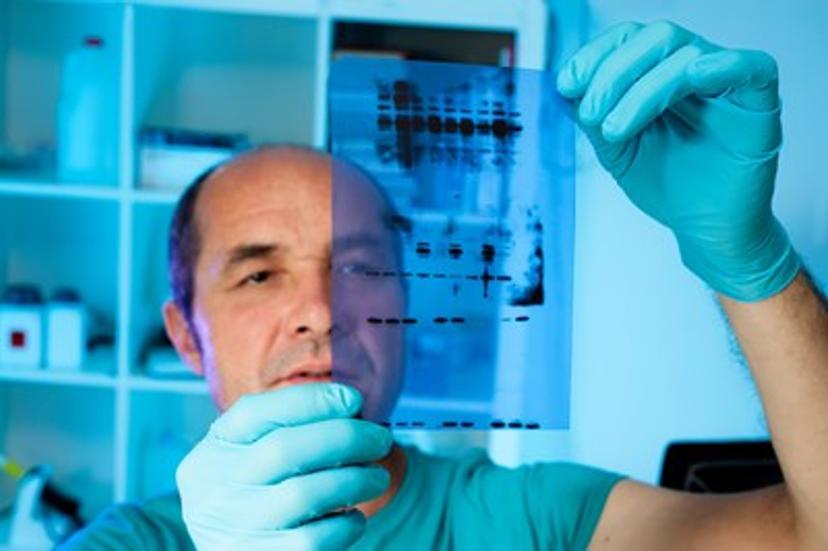
Western blot or immunoblotting is a rapid and sensitive technique that uses antibodies for the specific detection of proteins separated by polyacrylamide gel electrophoresis (PAGE) and immobilized onto a nitrocellulose, nylon or PVDF membrane. Western blot requires successive steps including transfer of the PAGE-separated proteins onto the membrane using either a wet or semi-dry system, pre-incubation on a blocking buffer that will help to reduce non-specific background signal, and incubation with a primary antibody that specifically binds the antigen of interest. Positive reactivity can be evidenced by the signal generated from a reporter enzyme or fluorophore conjugated to a secondary antibody that recognizes the primary antibody. The team at Rockland Immunochemicals routinely performs western blots for analysis of gene expression, antigens and antibodies. These are some useful tips to share for successful results:
1.Determine the best ratios of the target protein and primary antibody
Although general guidelines for protein loading and antibody dilution are recommended by the literature and antibody manufacturers, the relative abundance of the protein of interest as well as the titer of the antibody used sometimes require further optimization of those parameters. In general, 1 μg of purified protein or 10 μg of a mixture of proteins (i.e. lysate) containing the protein of interest should be enough to be detected by a solution containing 1 μg/mL of primary antibody. Nevertheless, visualization of low abundance proteins in a cell lysate might require as much as 50 μg of total protein and at least 2 μg/mL of antibody. Conversely, high background or undesired cross-reactivity can be modulated by adjusting these parameters in the other direction.
2.Keep up the protein transfer efficiency
In general, proteins can be successfully transferred by applying ~14V overnight in a wet transfer system or a maximum current of ≈0.8 mA/cm2 of gel area in a semi-dry system. Some proteins requiring improved transfer efficiency onto the membrane include large proteins exceeding 100 kDa or very hydrophobic proteins. They can be subjected to extended transfer times at high power using the semi-dry system but will require cooling to keep a constant transfer temperature of ~20°C. Also, the transfer buffer can be modified to increase transfer efficiency by adding SDS at a concentration of 0.1% (w/v). If using nylon membranes, SDS and methanol should not be used.
3.Anticipate the effect of gel thickness in western blot
Gel thickness has a double effect in immunoblotting, influencing both quantity and quality of antigen detection. In general, the thickness and acrylamide percentage of the gel inversely correlates with protein transfer efficiency and band diffusion, with gels 0.5 – 0.75 mm transferring more efficiently than thicker 1.0 – 2.0 mm gels. Also, the protein bands from thinner gels usually resolve better and provide crisper, well-defined detection.
4.Make sure to equilibrate membranes and gels on transfer solution
Always remember to equilibrate the membrane 10 to 15 min in transfer buffer before transfer and since PVDF membranes are hydrophobic and will not wet from just being placed into transfer buffer, first immerse 2s in 100% methanol, then equilibrate 10 to 15 min with transfer buffer. If the membrane dries out, wet once again with methanol and then transfer buffer. Following electrophoresis, it is recommended to equilibrate the gel 30 min at room temperature in transfer buffer to prevent a change in the size of the gel during transfer. Changes in gel dimension usually result in a blurred transfer pattern.
5.Cleaner blots with the right blocking solution
One of the most critical parameters to obtain clean western blots is the choice of an appropriate blocking agent. Blocking solutions work better when supplemented with a mild detergent like Tween-20, usually between 0.05% and 0.5% (v/v). A number of blocking agents can be used and these include immunoanalytical grade non-fat dry milk (blotto), BSA fraction V or normal serum at working concentrations ranging between 0.5% - 5%. Serum may be the best solution for very problematic backgrounds, apparently by reducing unspecific interactions between the primary antibody and the blocking agent. When using serum, it should be from the same species as the primary antibody or from the same species as the secondary when secondary antibody detection is used. Other applications including fluorescence detection should be performed using fluorescence dedicated reagents for optimal results.
6.Optimize your incubation time
The potency of a primary antibody might be leveraged by properly adjusting the incubation time with the antigen. In many cases, one hour incubation should be enough to visualize the protein of interest, however, overnight incubation at 4⁰C will allow enough time for the antigen-antibody reaction to occur and result in detection of a positive signal. One hour incubation at room temperature is usually enough for the conjugated secondary antibody and certainly, it should not be extended more than three hours since it might generate high background during detection.
7.Sometimes antibodies won’t recognize denatured proteins
Consider performing gel electrophoresis under non-denaturing conditions when you have your antibody working in other immunoassays but not in western blot. Since proteins are usually separated under denaturing conditions during gel electrophoresis, this restricts the detection of proteins by antibodies recognizing structural epitopes in non-denatured proteins.
8.Western blot of phosphorylated proteins
Some of the factors to consider when performing immunoblotting with phospho-specific antibodies are buffer compatibility, antibody specificity and protein abundance. The use of Blotto or other blocking mixtures containing dry milk is unsuitable because phospho antibodies could bind to a number of protein constituents in milk. Instead consider using BSA-based or alternative blocking buffers. A common problem related to phospho detection is that the phospho antibody is unable to detect the low abundant proteins of interest in a cell lysate. This problem can be overcome by means of immunoprecipitation as described in tip #9.
9.Detection of low abundance proteins
Detection of low abundant proteins by western blot can be achieved by immunoprecipitation of the target protein using a specific antibody, enabling more of the protein of interest to be loaded in the sample lane. Depending on the amount of lysate used in the immunoprecipitation, strong amplification of signal can be achieved by this method. Since the target protein can co-migrate with the heavy or light chain of the immunoprecipitating antibody that will react with the HRP-conjugated secondary antibody and obscure the signal from the protein of interest, it is necessary to use a qualifying reagent, i.e. TrueBlot, which avoids such interference and provides clear, unambiguous protein detection. Also, study of low abundant proteins by western blot can be aided by the use of enhanced chemiluminescence (ECL) that allows detection of pico and femto amounts of target protein.
10.Stripping and re-probing membranes
When the same membrane is required for testing of several proteins using different antibodies, stripping and re-probing is always possible although it might need to be empirically optimized for a particular assay since each antigen-antibody interaction is always distinct. In general, stripping buffers are reagents that combine low pH, detergents, reducing agents and/or heat in order to remove residual antibodies. Although repeated re-probing can lead to loss of signal, several re-probings are generally possible. Biotin-streptavidin interactions cannot be dissociated by this method but the whole complex can be removed away from the bound protein on the membrane.
Written by Camilo Moncada, PhD, Director of Quality Control at Rockland Immunochemicals. Dr. Moncada has performed research studies for more than 10 years and has been an active participant in several diverse projects that have resulted in publications in the areas of immunology, parasitology, cancer and lung disease. At Rockland, he applies his expertise on molecular and cellular biology, biochemistry and proteomics for antibody development and validation.