Western blotting expert shares the secrets to success in a GxP lab
See how high-quality imaging with sensitivity, resolution, and data security ensures confidence in your biomolecular research
19 Mar 2021

If you’re developing a new drug, cosmetic, or food product, you’re probably familiar with GxP. GxP – or good practice – guidelines define and track each stage of development and manufacturing to ensure that your final product not only performs its intended purpose, but that it’s also safe for consumer use. Having a GxP framework in place is becoming increasingly important – and data traceability, accountability, and integrity are at the heart of this.
“GxP is so very important today, especially for our customers in pharma and biopharma,” explains Sowmya Balachandran, Global Product Manager at Cytiva. “Good traceable data not only increases confidence in your final product but also helps you track and identify any issues should they ever arise.”
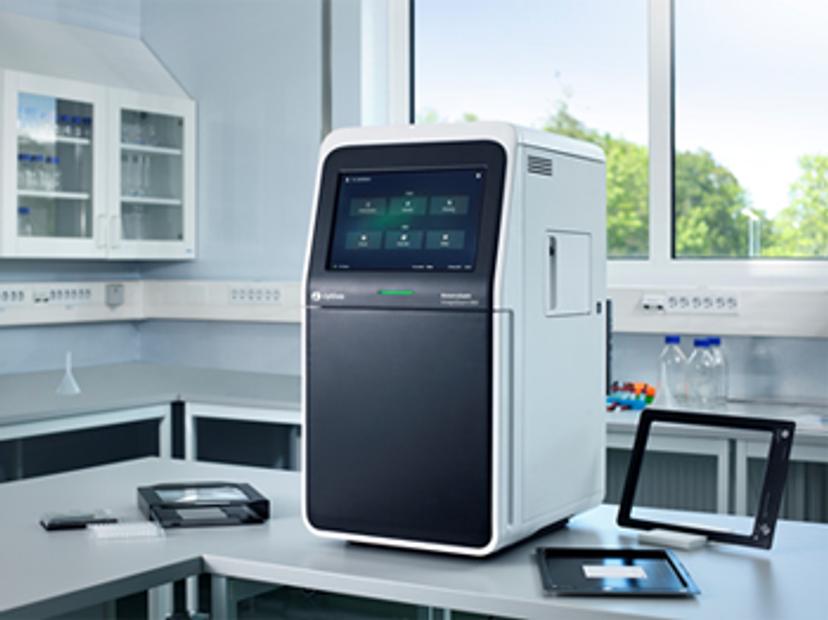
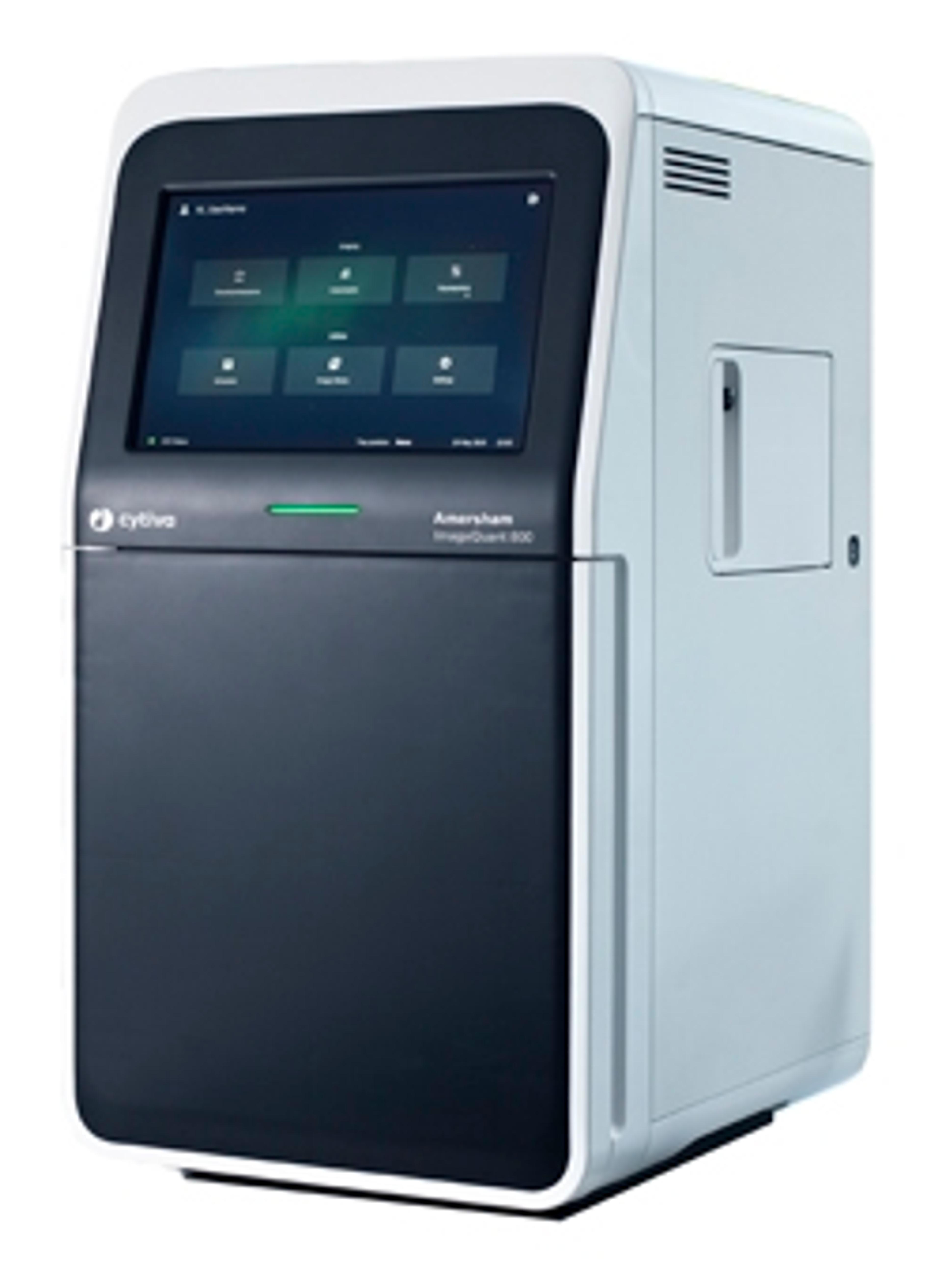
In this interview, Sowmya shares insight into the advancement of this fundamental technique for GxP-certified labs. She also explains how Cytiva’s Amersham™ ImageQuant™ 800 western blot imaging systems have a full suite of features aimed at supporting regulatory compliance.
Western blotting: A ubiquitous tool for biomolecule research
Unchanged for the past four decades, western blotting is a very simple, high-impact tool that helps answer some very basic but important research questions: Are there proteins present here? What proteins are present? How much of my target protein is present? As a result, western blotting has had a lasting consequence on workflows across research fields.
“It’s one of those underrated techniques because it's perhaps not the most exciting thing around to talk about, but it's one of those techniques without which we wouldn't have the answers that we have today,” says Sowmya.
Despite its simplicity, gel electrophoresis and western blotting is a somewhat fragmented process. From the initial sample preparation to final image analysis, each step requires different instruments, consumables, and reagents that often come from different suppliers.
This presents many challenges. Firstly, the success of a western blot depends on the user’s level of experience. If you can master western blotting as a skill, then you can get good results easily and cheaply – which is partly why many scientists rely heavily on this method. Then, after you’ve prepared a good blot, the next hurdle is making sure that you have access to an imaging system that can adequately detect target proteins for reliable downstream analysis.
“Anyone who's working on proteins will perform western blots and need to image them, so to be able to provide a product that almost every research lab can use feels amazing,” says Sowmya.
Sowmya Balachandran’s top tips for achieving GxP:
- Take a step back and look at the entire workflow. If there are multiple steps, introduce small checkpoints to close these gaps.
- Keep revisiting any processes to see if there are any improvements you can make to ensure data security and integrity.
- Remember, compliance depends on you, the user. Always use trusted suppliers and validate your workflow thoroughly.
From biomolecules to celestial bodies: Uncompromised, high-quality imaging
“If you're a skilled technician running a western blot, but your system can’t pick up the bands of interest and differentiate these against the background, then you’ll need to repeat the entire experiment. It’s time-consuming and unnecessary,” Sowmya says. This is especially true if you’re investigating a protein expressed in very small quantities. For effective biomolecule research, labs need imaging systems that can detect low-intensity signals and differentiate them from the background noise.
Traditionally, imagers tend to have a trade-off between resolution and sensitivity. To solve this, Cytiva developed the signal-to-noise optimization watch (SNOW) mode on its ImageQuant™ 800 imagers, which uses signal averaging – a well-known technique used in astrophotography to visualize distant stars.
SNOW mode works by capturing multiple images and continuously averaging the signal over time. This process reduces random noise while allowing the signal to remain constant, improving the signal-to-noise ratio. SNOW mode helps to enhance image quality with reduced noise and better resolution. Find out more about SNOW mode in this application note>>
“In terms of performance, the ImageQuant™ 800 hits that sweet spot where you can image difficult-to-see samples and simultaneously have the resolution that you need,” says Sowmya. “But what an industry lab needs more than just an imager that gives you good images is a layer of security for its data.”
Data security in the modern, electronic lab

The ImageQuant™ 800 GxP is a new module designed to help users maintain electronic data records in compliance with FDA 21 CFR Part 11 and EU GMP Annex 11 regulations. It includes features like operating system access control, audit logs, and image authenticity checks.
Today’s labs need to consider data security from various angles. If multiple people need access to the imager or its output, they probably need different levels of access. To address this, the ImageQuant™ 800 GxP lets you assign user groups that grant varying levels access to the required software functions.
The module also tracks and saves a full, time-stamped audit trail of everything that happens in the imager. But beyond imaging steps, it also has features to address larger, fragmented workflows.
“You often start with a sample prep stage somewhere on your lab bench. Then you perform electrophoresis, followed by western blotting, and then you finally reach the imaging step – where you put the blot through your imager before looking at the image file in your analysis software. That’s when the file leaves the secure environment of the imager,” Sowmya says. “Normally, users record an experiment ID or a sample ID in their electronic notebook at the start of the experiment, but there needs to be a way to seamlessly link up your sample, the imager, and the final analysis steps.”
To link up this fragmented process and ensure consistent electronic reporting, the ImageQuant™ 800 GxP uses a string of “digital handshakes” to connect all stages of the workflow. It uses a mandatory image ID field that helps users trace experiments from the origin of the sample, through imaging, to the final analysis output.
Each sample gets an assigned ID that you can enter into the imaging system. This ID connects each sample to its first step, along with everything that has happened previously. Additionally, to reduce the risk of tampering before analysis, images on the ImageQuant™ 800 GxP carry a special security tag within the metadata. The ImageQuant™ TL 10 GxP analysis software reads this tag and alerts you to any suspected tampering – and it won’t open tampered images to better ensure data security.
Future innovations responding to scientists’ needs
Another key factor for success is validation. To ensure scientists have confidence, Cytiva provides access to a validation support file on their regulatory support page, which includes full development documentation, change control notifications, and external assessment reports.
“The self-checklist inside our validation support file documentation helps users check and validate their workflows for compliance," Sowmya explains. “We also went a step further with an external audit that looked at how we developed the ImageQuant™ 800 GxP, what the features are, and checked all of our system documentation from a GxP perspective. Customers can access this audit report along with the certificate by downloading the validation support file on our website.”
Looking to the future, Sowmya and the team at Cytiva understand that there is always room for improvement. “We are committed to offering the best solution and products to our customers,” concludes Sowmya. “Our product development process is based on customer feedback. This process continued after launch and we always aim to improve our offering, so our customers can always be confident that they have the best-in-class solution with Cytiva.”
Cytiva's vision:
Cytiva envisions a world in which human health is transformed through access to life-changing therapies. Its products and services aim to support customers in helping advance and accelerate therapeutic development. Its imaging products are used in basic research – including western blotting and gel electrophoresis – to help identify, characterize, and quantify proteins, which is often the first step in any research workflow and serves as a basis for more advanced experiments that eventually lead to a better understanding of diseases and the effect of drugs on cells.
Do you use Cytiva products in your lab? Write a review today for your chance to win a $400 Amazon gift card>>

