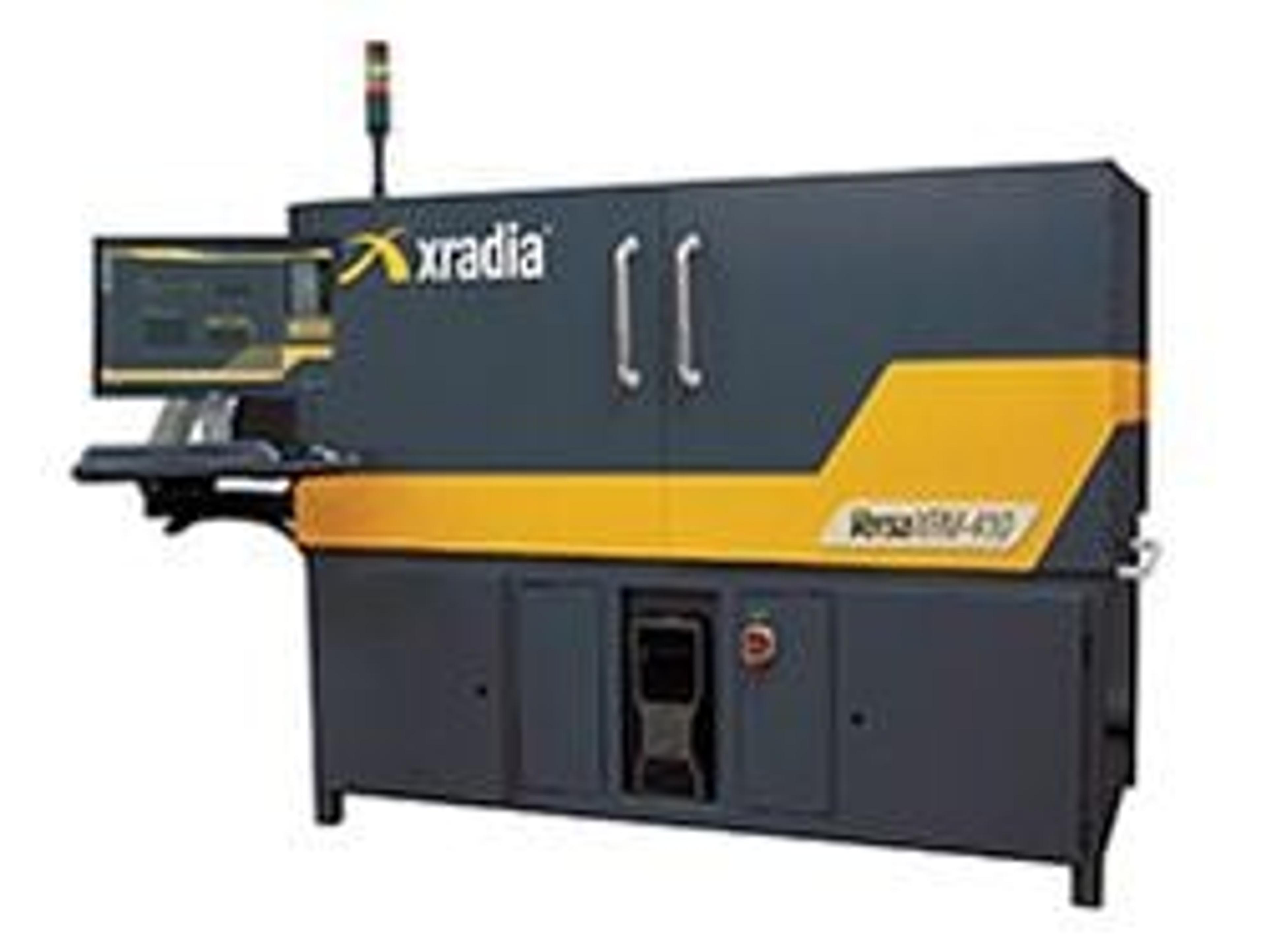
Xradia Announces VersaXRM-410 to Bring Revolutionary X-ray Microscope Technology to More Researchers
13 Jun 2013Xradia, Inc. is announcing the expansion of its lab-based VersaXRM family to bridge the gap between high-performing 3D X-ray microscopy (XRM) solutions and traditionally lower-cost, less capable projection-based computed tomography (CT) systems. The VersaXRM-410 delivers the advantages of the VersaXRM family including highest resolution and contrast and in situ capabilities that enable ground-breaking research for the widest range of sample sizes.
The University of California, Irvine, is using the VersaXRM-410 to nondestructively characterize the microstructure and mechanics of composite materials with applications in civil, mechanical, aerospace and biomedical engineering. Professor Lizhi Sun says, "The newly installed VersaXRM-410 lets us characterize the behavior and local deformation of materials in 3D in their native environments (in situ) while uniquely maintaining sub-micron resolution across an array of sample dimensions and environments. What's even more powerful is that we can extend the understanding of a material's microstructure to the 4th dimension (3D + time) by studying how a microstructure evolves over time, and quantify that change. Only non-destructive X-ray tomography lets us achieve that goal."
Dr. Kevin Fahey, Chief Materials Scientist and VP of Marketing at Xradia, says the VersaXRM family was architected to make advanced imaging capabilities available to more researchers worldwide. "Research facilities face economic constraints, but at the same time, studies increasingly demand the non-destructive, high-resolution, high-contrast 3D imaging enabled by XRM," Fahey says. "VersaXRM brings synchrotron-like capabilities to the lab, overcoming the resolution and contrast limitations of traditional micro-computed tomography approaches to advance studies being conducted today and into the future."

