ZEISS Hosts 5th Annual Light Sheet Fluorescence Microscopy Workshop
22 Oct 2013Over forty international scientists gathered for the 5th Annual Light Sheet Fluorescence Microscopy Workshop in September in Thornwood, New York. The meeting was held at the U.S. headquarters of Carl Zeiss Microscopy, LLC. Researchers presented data and shared ideas regarding instrument development, standards, applications and specific applications of light sheet fluorescence microscopy (LSFM) and related technologies to various biological model organisms such as zebrafish, drosophila, mouse and others.
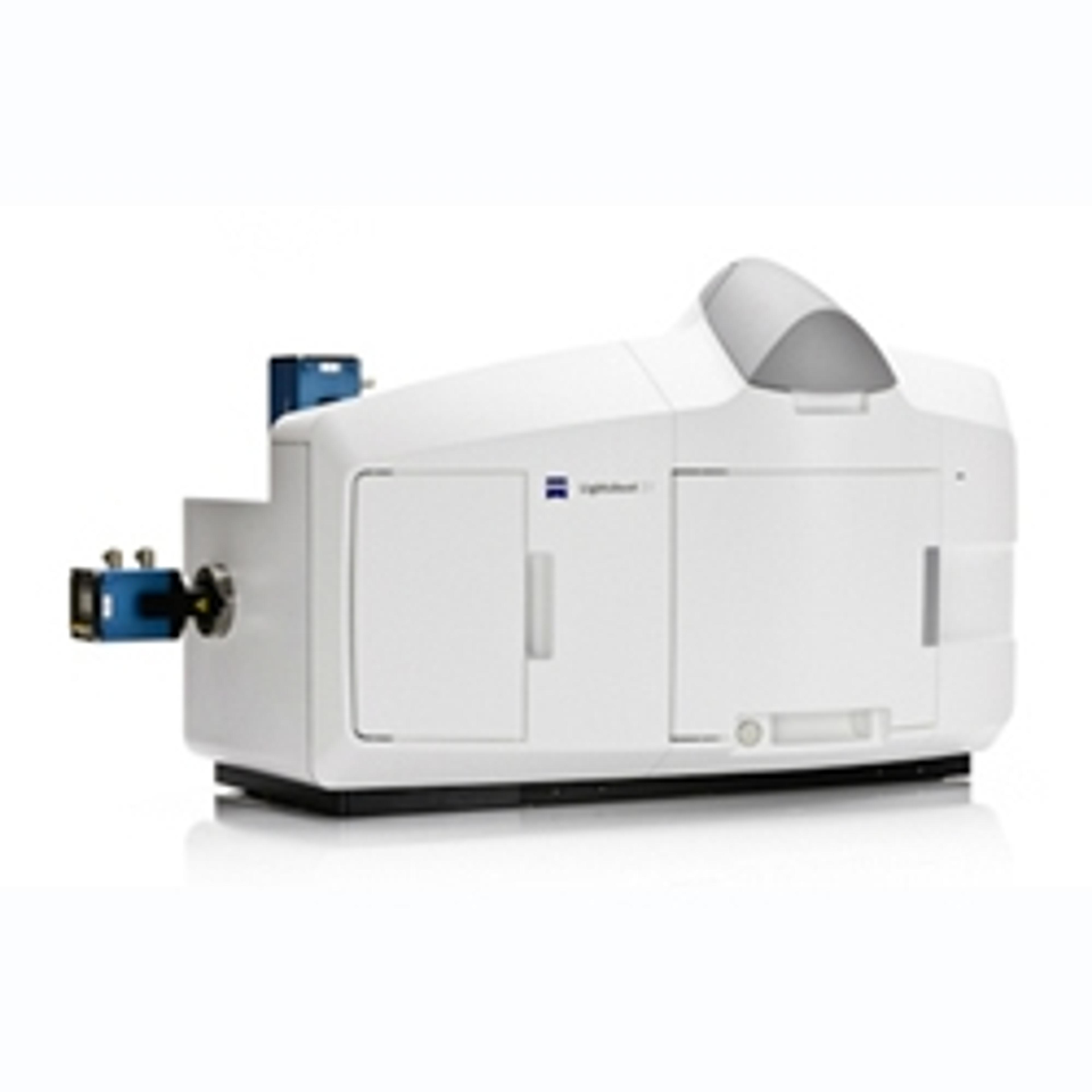
The LSFM community was formed in 2009 by the bioscientists Pavel Tomancak, Emmanuel G. Reynaud and Peter Santi as a forum for developers and early adopter users of light sheet fluorescence microscopy. This technology combines optical sectioning with Multiview imaging to observe tissues and living organisms with impressive resolution. LSFM minimizes the illumination light dose on the specimen by selectively irradiating only the observed area and thereby virtually eliminates both phototoxicity and photobleaching compared to other optical sectioning techniques, such as laser scanning confocal. This makes light sheet fluorescence microscopy the most gentle fluorescence imaging tool for living specimen and enables long term imaging of, e.g., entire, developing embryos of, for example, drosophila or zebrafish embryos. The ability to collect three dimensional images of such large samples extremely quickly provides new insights in the dynamics of developmental processes and increases research productivity.