ZEISS Introduces New Technology for 3D Superresolution Microscopy
18 Nov 2013Expanding its 3D microcopy portfolio, ZEISS introduced superresolution photoactivated localization microscopy (PALM) in 3D at the Society for Neuroscience Annual Meeting in San Diego, California.
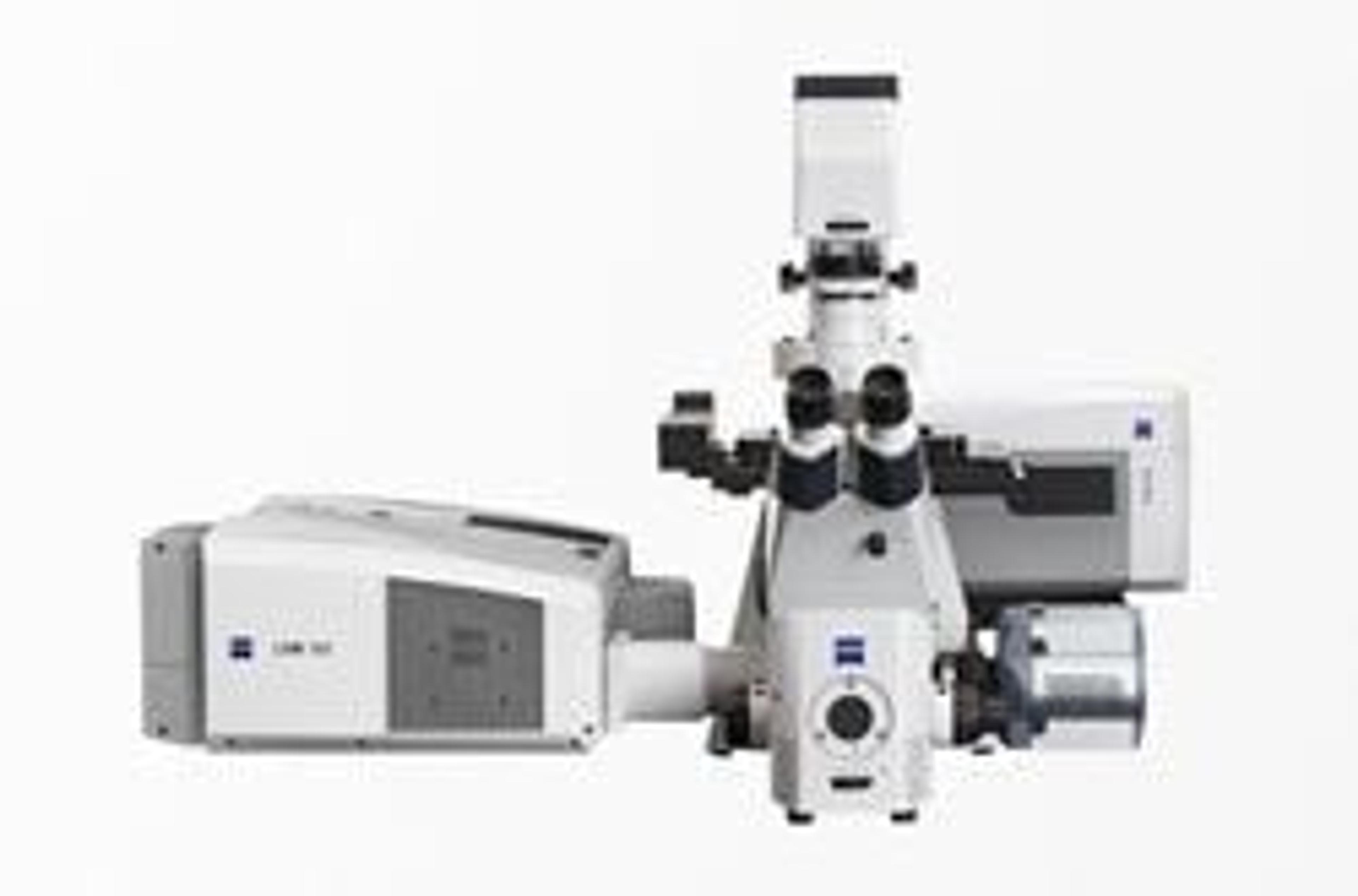
With a new module, ELYRA P.1 now enables superresolution photoactivated localization microscopy (PALM) in 3D for endogenously-expressed photo-switchable fluorescent proteins. Users can capture highly resolved structures in 3D, while treating the sample so gently it stays fit for long-term observation.
In PALM, photo-switchable fluorescent molecules are sparsely activated so that only one out of many will be in its on-state within a single point spread function (PSF). In 3D, the PSF shape codes for the z-position. The localizations are plotted in a new image to create the super-resolved image. ELYRA P.1 achieves resolutions in the range of 20-30 nm laterally and 50-80 nm axially.
“With ELYRA, researchers can investigate the structural arrangement of one or multiple proteins, reveal the ultrastructure of cell organelles in 2D and 3D as well as map and count molecules within a structure. Sophisticated algorithms relate photon statistics to precision information in all directions, so researchers can display their structures fully rendered in 3D”, explains Dr. Klaus Weisshart, Product Manager for ELYRA at ZEISS.
Users can select the right tool for their application: Superresolution structured illumination microscopy (SR-SIM) or PALM. ELYRA PS.1 combines both technologies in one system. The ELYRA system can be integrated with the ZEISS laser scanning microscopes LSM 710 or LSM 780. Plus ELYRA works seamlessly together with ZEISS scanning electron microscopes in a correlative workflow allowing scientists to navigate between information from different imaging modes via ZEN Shuttle & Find.

