ZEISS to Offer Advanced Microscope Technology for Imaging of Cleared Tissue
14 Nov 2014
At the annual Neuroscience meeting in Washington, D.C., November 15-19, 2014, ZEISS will present new microscope hard- and software for imaging of cleared tissue.
Clearing methods make tissue transparent, thus allowing scientists to image deep into large biological samples such as tissue sections, brains, embryos, organs, spheroids or biopsies. The enhanced optical penetration depth even allows detection of fluorescent signals from whole organs. This means clearing is a promising technique when, for example, investigating neuronal networks in mouse brains.
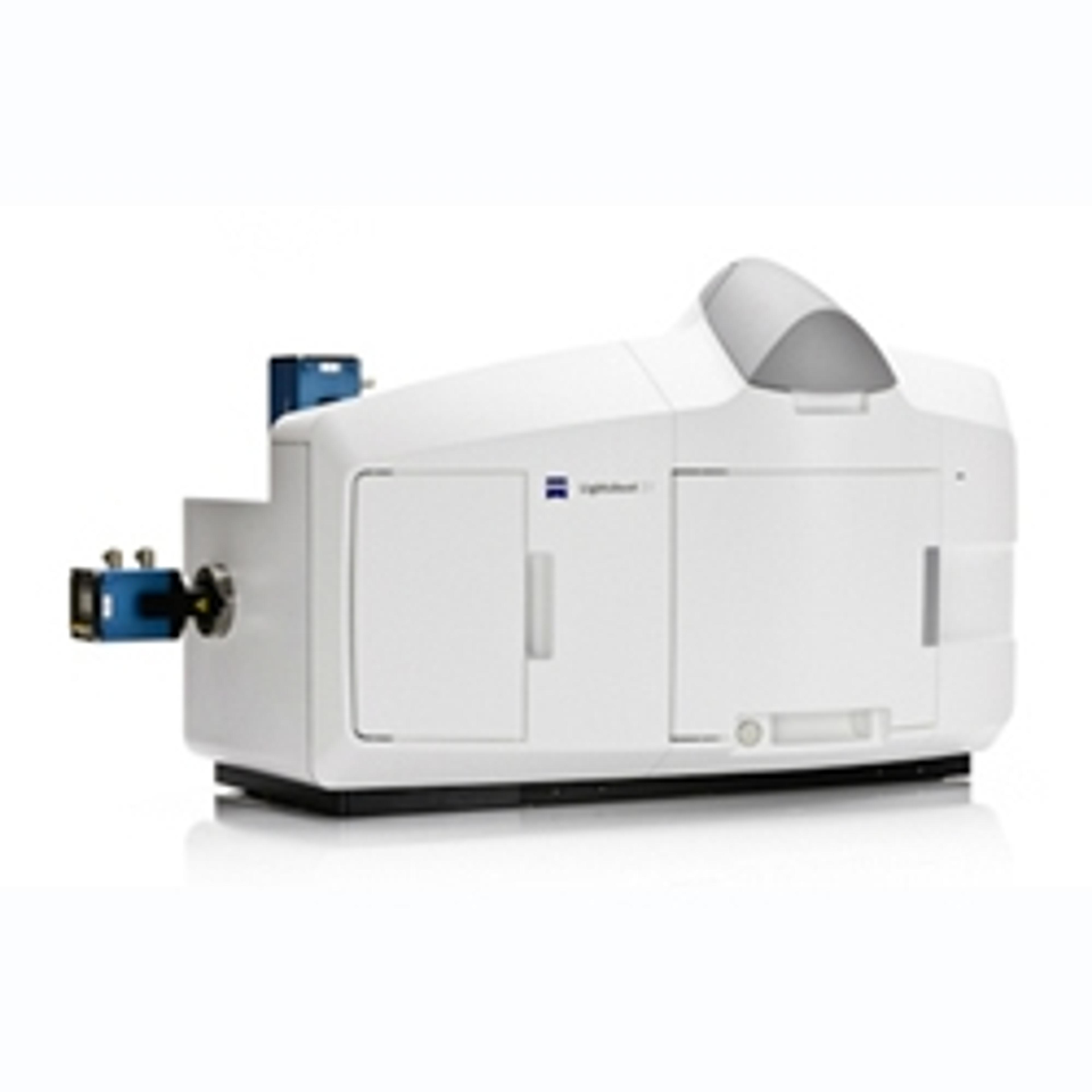
The microscope system ZEISS Lightsheet Z.1 combines the advantages of clearing with light sheet fluorescence microscopy. Researchers are now able to image large cleared specimens with high light efficiency, speed, and almost no photo damage. ZEISS Lightsheet Z.1 acquires multiple tiles of Z-stacks with several thousand high quality images. A typical imaging speed of 10 to 40 frames per second reduces the imaging time from hours to minutes.
ZEISS now offers a range of objectives with long working distances, adapted to the refractive index of respective clearing media. In addition, the sample holder of ZEISS Lightsheet Z.1 is now ready for the special requirements of large and cleared samples.
3D imaging of large samples produces datasets in the Terabyte range that need to be stored, transported, viewed, and analyzed. ZEISS has worked together with arivis AG to provide a software platform that addresses these needs during the complete workflow. The imaging platform arivis Vision4D is a modular software package for fast visualization of 2D, 3D and 4D images of nearly unlimited size independent of RAM.
Image: Thy1-EGFP M-line mouse hippocampus, optically cleared in LUMOS clearing agent, imaged with ZEISS Lightsheet Z.1, data processing and 3D rendering in arivis Vision4D software. Sample courtesy of O. Efimova, National Research Center Kurchatov Institute, Moscow, Russia.

