Microenvironment and 3D Image Analysis Key in the Development of More Efficient Drugs Against Complex Diseases
Learn about the technologies advancing phenotypic screening at HCS Pharma
14 Oct 2018
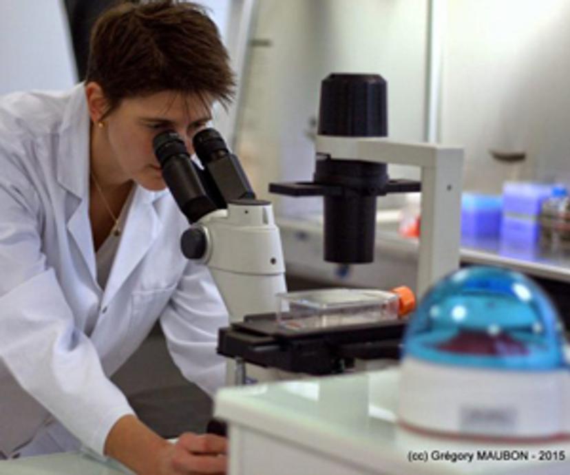
Nathalie Maubon, Ph.D., launched HCS Pharma in 2014, inspired by an idea: to obtain and select effective drugs, more relevant cellular models are needed for screening.
The biotechnological start-up is focused on in vitro preclinical research and development with a specialization in cellular imaging: high-content analysis (HCA) and high-content screening (HCS).
In this SelectScience® interview, Maubon provides insight into the development of in vitro models and assays to advance phenotypic screening, and the cellular imaging technology enabling her work.
SS: Tell us about your role at HCS Pharma
NM: I am the CEO and CSO at HCS Pharma. I manage the company by surrounding myself with a young, dynamic and efficient team. Altogether, we develop new in vitro models and assays by taking into account the cellular microenvironment, because it plays a fundamental role in cell homeostasis and in pathological conditions. To reach this goal, we have recently acquired BIOMIMESYS® technology, which is a unique and natural hyaluronic acid-based hydroscaffold, biofunctionalized with other extracellular matrix (ECM) components to better mimic the microenvironment of every organ. With this technology, we develop healthy or pathological mini-organs, by mixing different cell types and cultivating them in our 3D matrix to get closer to in vivo situation. These new 3D cellular models are tested in phenotypic screening.
I am really convinced that to find effective drugs, we must reproduce the pathology as a whole, taking into account many (if not all) the cells of the diseased organ and their microenvironment.
Nathalie Maubon, Ph.D. HCS Pharma
SS: Why is phenotypic screening important to drug discovery? What can you measure with phenotypic screening that you cannot with other methods?
NM: So far, in vitro screening assays (HTS) have been made extremely simple to allow chemists to perform QSAR (quantitative structure-activity relationship) studies. HTS is used to identify molecules that are active on a particular molecular target (for example, a receptor). However, many diseases are complex/multifactorial conditions like cancer and neurodegenerative diseases. They are caused by many contributing factors and affect different cellular populations; therefore focusing on a single target (or a single cell type) is not enough. To get more effective drugs against such pathologies, the biological assays used for screening should be multi-target and multi-cell types. Phenotypic screening by cell imaging allows such assays.
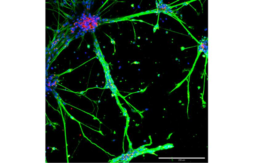
I am really convinced that to find effective drugs, we must reproduce the pathology as a whole, taking into account many (if not all) the cells of the diseased organ and their microenvironment. For instance, inflammation is known to play a role in many conditions: the co-culture of the target cells and immune cells is needed to increase the relevance of the cellular models related to these diseases. High-throughput cell imaging allows us to characterize many parameters in a single experiment: the number, size and morphology of the cells, together with cellular stress, organelles’ modifications or target expression/localization.
SS: How have 3D cell models helped you to advance your work?
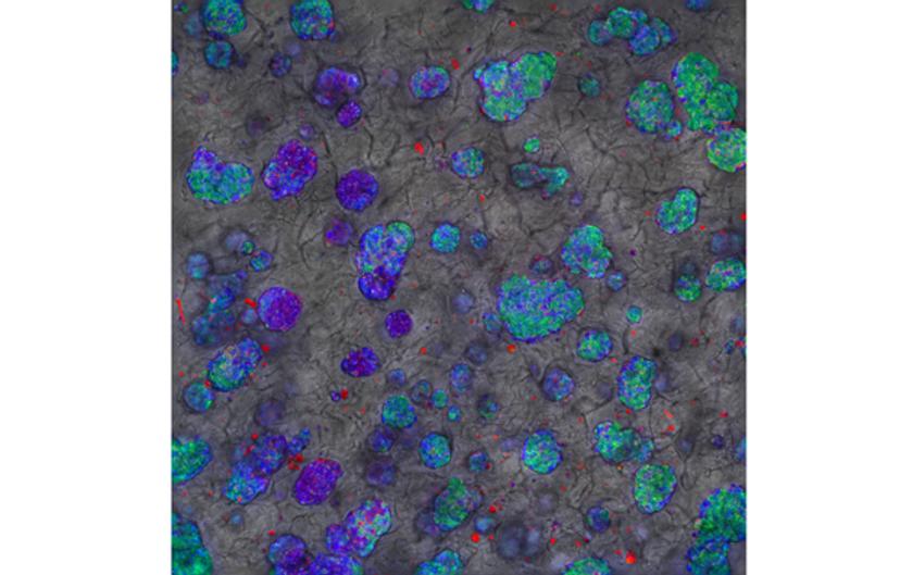
NM: 3D technologies including ECM, such as BIOMIMESYS® hydroscaffold, help to improve the relevance of our cellular models in terms of structure and function. 2D culture is linked to a rapid dedifferentiation of the cells. For example, primary hepatocytes cultivated in 2D culture — even if they are cultivated in a sandwich of collagen — keep their functionality for a maximum of seven days. When we cultivate primary hepatocytes in BIOMIMESYS® Liver (which mimics the ECM of this organ), we keep them alive and functional for a month. We obtained similar results with other cells, such as adipocytes with BIOMIMESYS® Adipose tissue. Furthermore, we observed different cellular behaviors or different morphologies depending on the ECM composition: it can be modulated to mimic a “pathological” matrix, for instance. We could see an increase in the proliferation of cancer cells when the rigidity of the microenvironment increased, as observed in tumors of higher grades. Thus, I am convinced that cultivating the cells in a physiological or pathological-like microenvironment, both in terms of composition and physical properties, is essential for developing new relevant cell models for screening purposes.
SS: What are the main challenges in phenotypic screening?
NM: The main challenges in phenotypic screening are the biology, and the image/data visualization and analysis in 3D.
First of all, developing relevant 3D cellular models is crucial to find new drugs which have higher efficacy and safety, as explained previously. Nowadays, HCS allows multiplexing up to four different fluorescent markers (blue, red, yellow, green). It is necessary to choose the markers that are followed for an experiment (morphological markers, markers of cellular stress, or proteins of interest). Making cellular models more complex by co-culturing cells requires the use of markers to identify each cell type and thus reduces the possibility of marker tracking.
Secondly, the use of 3D cellular systems requires image stack acquisition in z direction. This increases the number of images to be taken during a screening, and therefore enhances image acquisition. The penetration of light in such systems being more difficult, imaging deep into cellular 3D structures is problematic: either the field of investigation is reduced, or the structures must be submitted to tissue clearing.
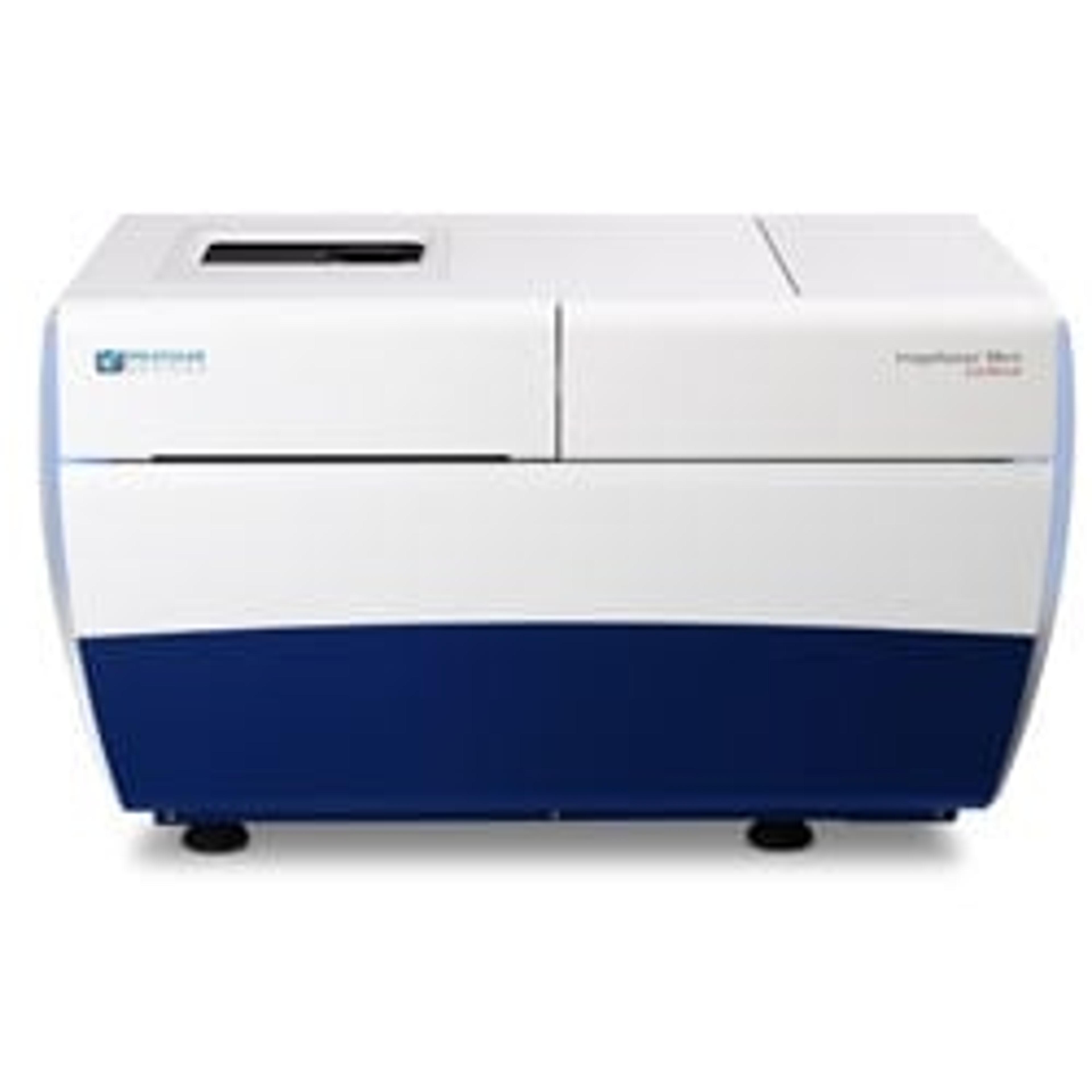
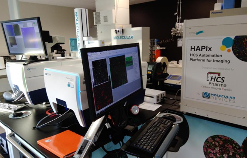
Thirdly, the images and data visualization and analysis are very challenging in 3D. Volume measurements must be done by taking into account three dimensions during image analysis. Visualization of 3D-reconstructed images by using virtual reality technologies might represent a solution. In HCS Pharma, we are working to implement this technology on Z-stack images that we have acquired using our two ImageXpress Micro Confocal High-Content Imaging Systems from Molecular Devices. These imaging systems form an integral part of our automated platform. Furthermore, Phenotypic screening is often a comparison between two (treated vs untreated) populations, in a supervised manner. Some treatments can modify the cells in a subtler manner than dead/alive, and more than two phenotypes can be observed. To go deeper into these processes, we would like to clusterize the compounds depending on the different phenotypes they induce. In this frame, HCS Pharma is also working on the establishment of data analysis procedures in a non-supervised manner by machine learning / deep learning and/or artificial intelligence.
SS: How is your partnership with Molecular Devices helping you to address these challenges?
- Which features of the ImageXpress Micro Confocal System benefit your work/results?
We acquire images in stacks in three dimensions using our ImageXpress Micro Confocal systems. The different pinhole sizes for confocal microscopy allow us to choose the best acquisition time / resolution ratio. Image acquisition is often the bottleneck in phenotypic screening. With our two ImageXpress Micro Confocal systems on our robotic platform, we are doubling our imaging capacity and thus our screening capacity. Furthermore, some options in MetaXpress allow us to analyze Z-stack images in 3D, in volume rather than area only.
- What are the goals of your partnership with Molecular Devices and how are you working together to advance the phenotypic screening services that you provide?
The main goals of our partnership are, first, to communicate together on phenotypic screening and, secondly, to learn together about the needs to go further in phenotypic screening. Mimicking pathological organs in 3D culture including the microenvironment and using microfluidic systems will help in the discovery of more efficient drugs against complex diseases. To go further, we need to work on biological systems, develop new imaging systems and innovative devices for a robotic platform to acquire images deeper and faster in a 3D biological system, and probably, in the future, include microfluidic systems. Other software should also be developed for image visualization/analysis in 3D (using virtual reality, for example). For the future of phenotypic screening systems, we are willing to work closely with Molecular Devices on these aspects.
SS: What do you see for the future of phenotypic screening in terms of applications and technology?
NM: For most biological assays, the issue about the most relevant cell system still remains unresolved. Many efforts are now being made to avoid the use of cancer cell lines, to better mimic the physiological/pathological environment and the cellular responses to pharmacological treatments. In this regard, the emergence of human induced pluripotent stem cells (iPSCs) is a first step in the design of physiologically more relevant screening models, in order to answer basic as well as disease-relevant questions. Moreover, the possibilities to model monogenic diseases using iPSCs will be expanded by genome editing with RNA-guided nucleases, like the CRISPR/Cas9 system: it enables the specific introduction of mutations in wild-type iPSCs or their correction in patient-derived iPSCs.
But an organ is not made only of cells. The cells are surrounded by an extracellular matrix (ECM) that differs from one organ to another. This ECM is modified in some pathologies and modifies cell behavior and homeostasis. To better mimic a pathologic organ (as tumor or fibrotic organ), the cells must be cultured in 3D in a microenvironment that corresponds to it.
I believe that dynamic 3D cell models including microenvironment represent the future of phenotypic screening, in parallel with the development of microfluidic platforms adapted to HCS. Microfluidics is the technology of manufacturing microminiaturized devices containing chambers and tunnels through which fluids flow. Microfluidic devices can reduce reagent consumption, and help to integrate and automate multiple assays (known as “organ-on-a-chip”, “lab-on-a-chip”). Facilitating imaging and tracking within these systems will for us be useful in phenotypic screening.
SS: How are you using augmented reality, and do you think this is something that we will see more of in the life science industry?
NM: Augmented reality (AR) is only a technology, very useful to “manipulate”, as naturally as possible, digital data: therefore, it can be used for many purposes. In HCS Pharma, we used AR to validate the conception of our HAPIx platform. Since we are focused on 3D culture and we acquire lots of pictures, it important for us to visualize results and to share them between colleagues and with customers. For example, AR on our scientific posters can help to better explain our results. It’s very simple and efficient.
We believe that the use of immersive technologies will emerge in life sciences. It is, in fact, a global trend in all industries. If you are interested, please visit our website dedicated to this topic. Feel free to contact us to exchange ideas about immersive technologies!