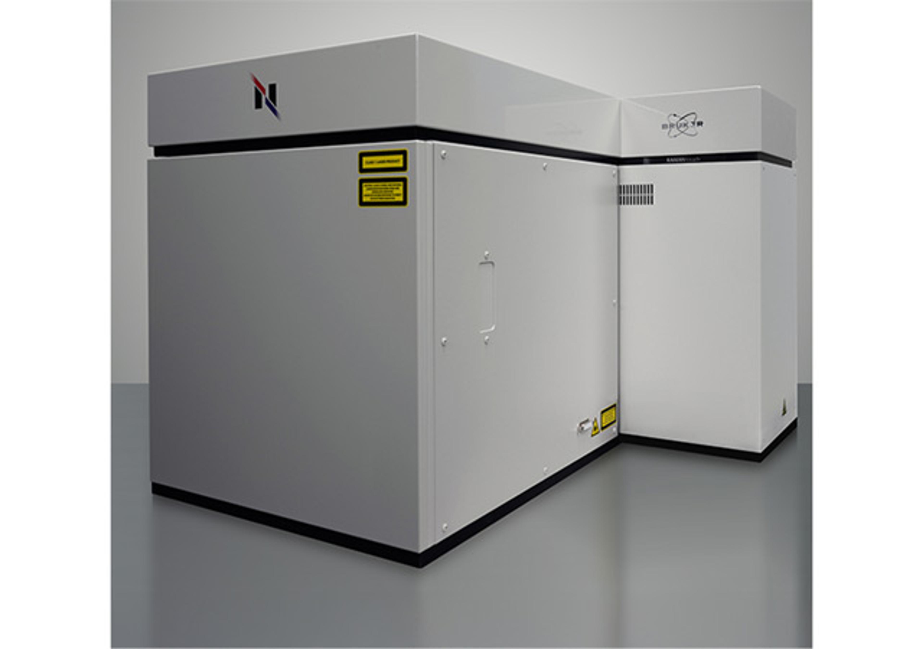
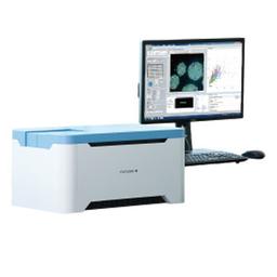
CellVoyager CQ1 Benchtop High-Content Analysis System
Compact footprint, lightweight bench-top device; no need for a darkroom.
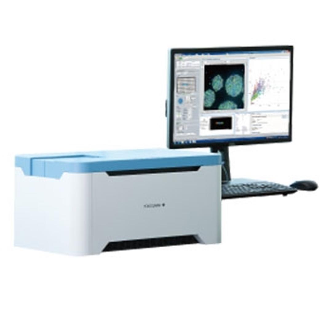
CellVoyager CQ1 Benchtop High-Content Analysis System


The supplier does not provide quotations for this product through SelectScience. You can search for similar products in our Product Directory.
Great instrument for the money!
Drug discovery
Excellent bench top confocal high content instrument with an easy to use interface. Exceptional value for the engineering and feature set.
Review Date: 30 Dec 2021 | Yokogawa Corp. of America
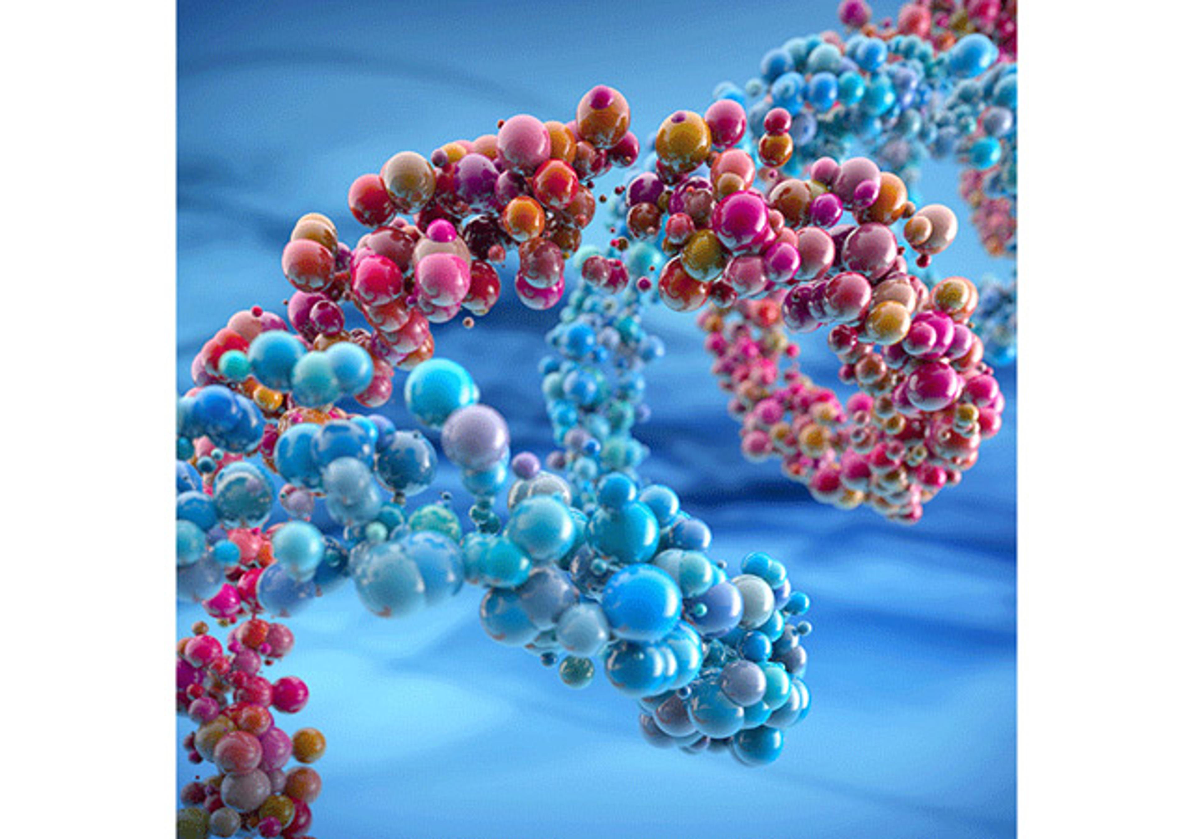
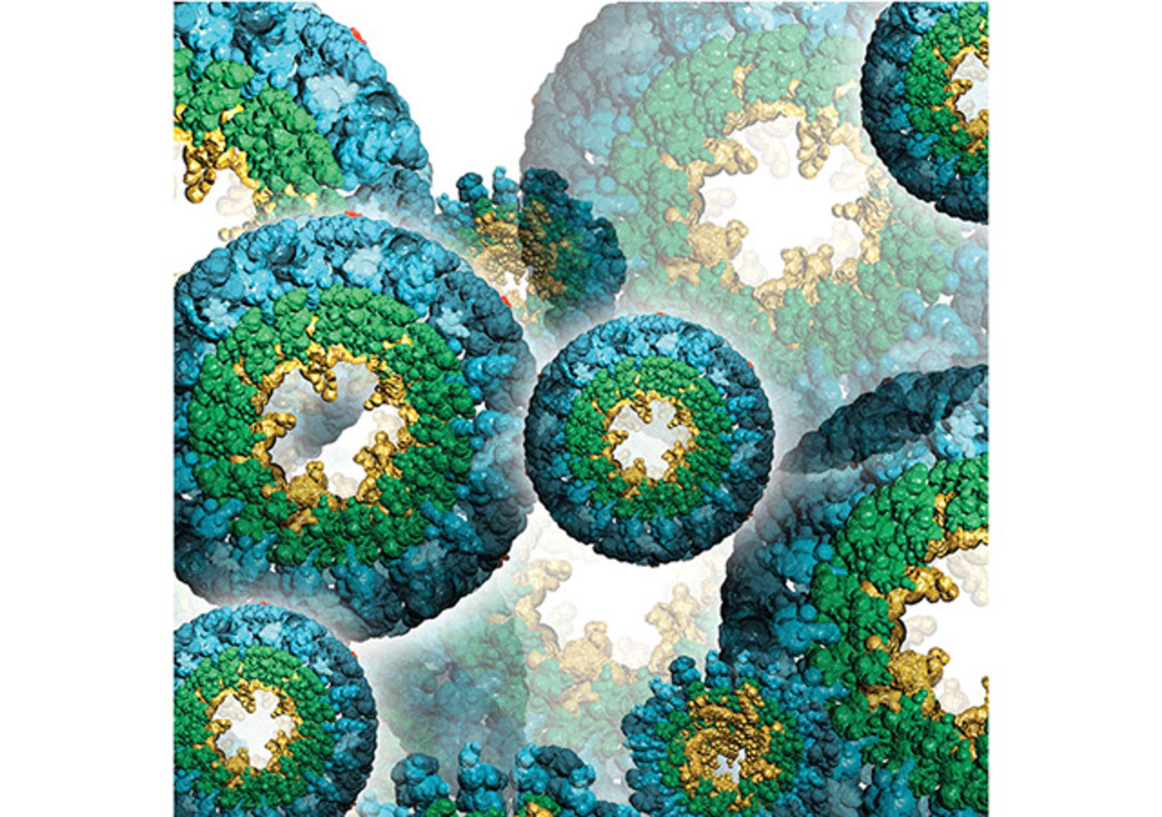
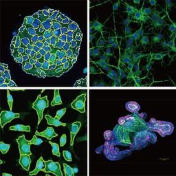
CellVoyager CQ1 enables 3D imaging and quantification of live cell clusters, such as spheroids within a 3D culture vessel, as they are, keeping the cells intact. CellVoyager CQ1 exports feature data in general formats which are readable by various third-party software for advanced data analysis. It is possible to construct a fully customized CellVoyager CQ1-based system by integrating with external systems, via robot for culture dish handling.
Enables measurement of spheroids, colonies, and tissue sections
- No need to remove cells from the culture dish, in contrast to traditional flow cytometry
- Nipkow spinning disk confocal technology allows high-speed yet gentle 3D image acquisition
- Rich feature extraction to facilitate sophisticated cellular image analysis
- Wide field of view and tiling capability enables easy imaging of large specimen
Enables analysis of time-lapse and live-cell
- High precision stage incubator and low phototoxicity of our confocal makes the analysis of time-lapse and live-cell are possible
- Max.20fps option for fast time lapse
- CQ1
High-quality image and similar operability to a traditional flow cytometer
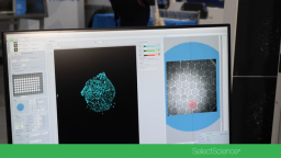
- Feature data and statistical graphs displayed in real-time with image acquisition
- Usable high-quality image as confocal microscope image.
- Easy to trace back to the original image from a graph spot, and make repetitive measurements
Open platform
- Connectable with external systems via handling robot
- Expandable to the integrated system as image acquisition and quantification instrument
- FCS/CSV/ICE data format readable by third-party data analysis software
- A variety of cell culture and sample dishes are applicable