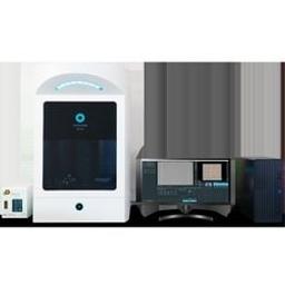
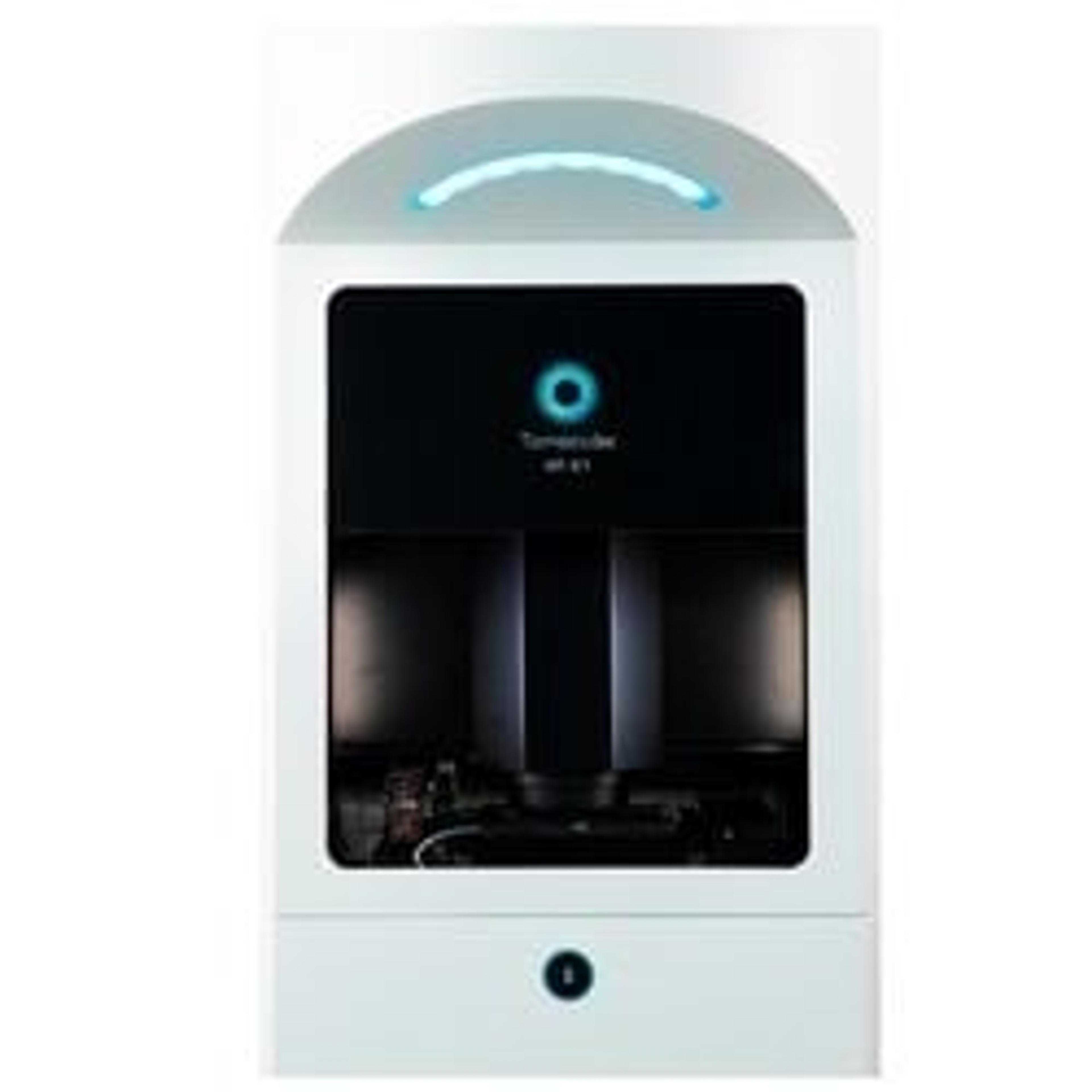
HT-X1
X-tra: Next-generation holotomography for high-performance imaging at scale
The supplier does not provide quotations for this product through SelectScience. You can search for similar products in our Product Directory.
Easy to use!
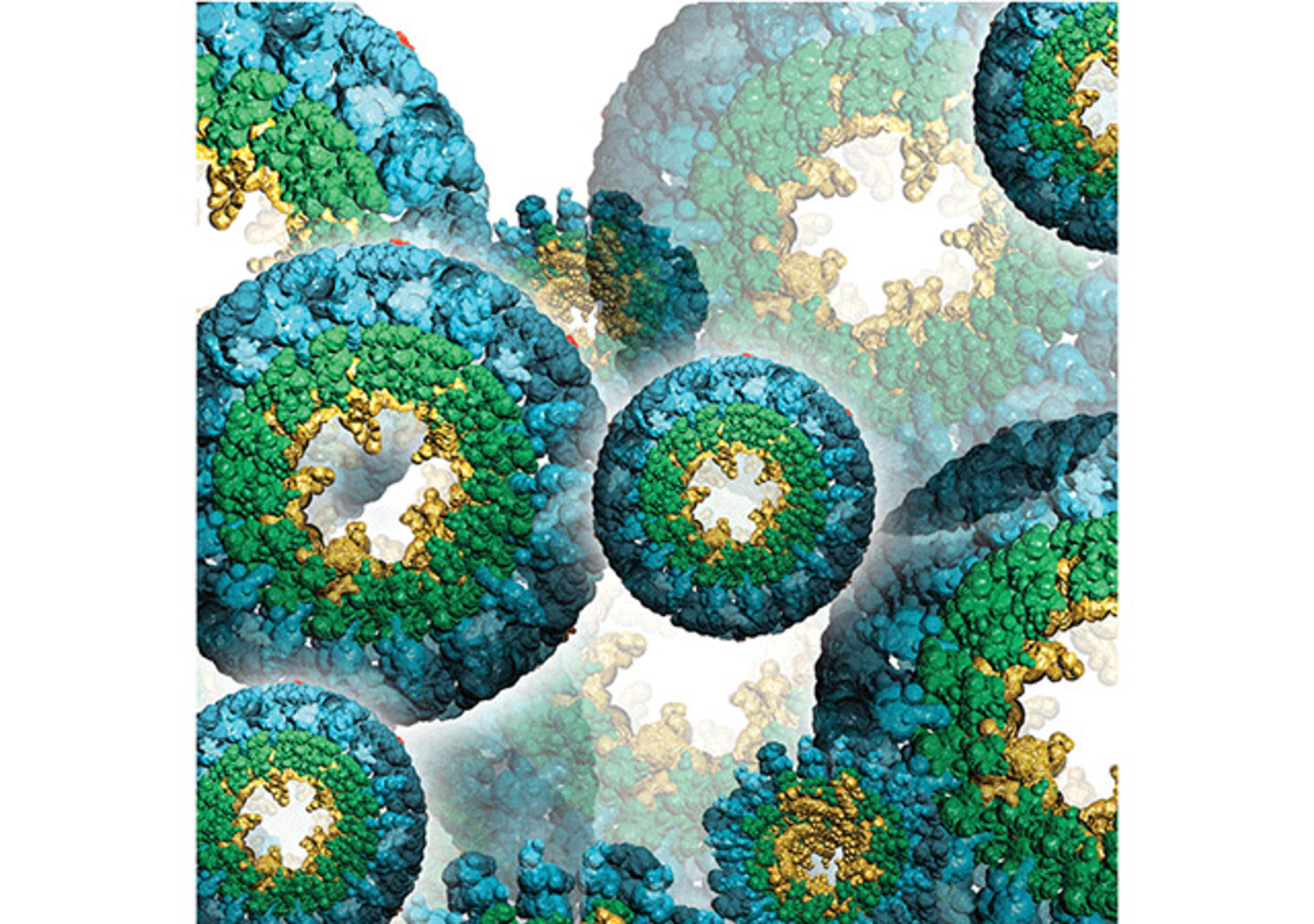
Analyze gas vesicles In microbes
I had the opportunity of demoing the Tomocube at Woods Hole. Such an easy to use instrument and the tech support is great. Label-free imaging is super cool.
Review Date: 21 Jul 2023 | Tomocube
So happy that I can use this machine for my experiment
Observing cell dynamics and lipid droplets
Such convenient but so powerful for observing cell dynamics. The incubator system is so stable that users can observe cells for a long time. Also, the company is always challenging to develop many novel tools for users to enjoy/explicit new aspects of HT-X1. Anyone can handle it, anyone can make amazing results
Review Date: 20 Apr 2023 | Tomocube
Tomocube HT-X1 is genuinely outstanding microscope
Cell imaging, Exosome imaging, Organoid imaging
If you are involved in cell imaging, the Tomocube HT-X1 would provide a fantastic experience. I have used various types of microscopes, including confocal microscopes. Based on my experience, I believe that the Tomocube HT-X1 is genuinely outstanding. The label-free images captured by theHT- X1 exhibit an astonishingly high level of resolution. I've been taking images of nano-sized exosomes and millimeter-level organoids, and I am getting satisfactory data!!! This microscope is highly user-friendly and optimized for capturing multi-tile images and time-lapses. Additionally, the engineering team is extremely dedicated and responsive to issues that arise during use and to my needs, striving to improve the product. I am delighted with this instrument.
Review Date: 29 Mar 2023 | Tomocube
Mothership equipment for cell imaging
Multimodal imaging of cells
Tomocube is an impressive system, setting itself apart as a unique and unparalleled solution in cellular imaging. It generates 3D images of cells in a phase contrast mode (hologram) and also acquires fluorescent images. The spatial resolution of the hologram is stunning and provides a wealth of information on cellular composition. In addition, the system supports automated high-throughput screening and live-cell imaging capabilities, making it a comprehensive and efficient tool for a wide range of cell experiments. Our team has utilized the Tomocube system extensively for various cell imaging experiments, including 2D culture and organoids, and we have been satisfied with the results. The user interface is also very clean and intuitive, and the customer support is outstanding.
Review Date: 29 Mar 2023 | Tomocube
Excellent product
Biology, Material science
Tomocube X1 is an excellent label-free imaging micrcoscope that open up a lot of novel findings in biology. It is very easy to use instrument and software. After we purchased, they support us very kindly and help us what we need.
Review Date: 28 Mar 2023 | Tomocube
The most valuable imaging system for live cell imaging!
Live cell imaging
The Tomocube's HT-X1 is the only imaging machine to observe the Adipogenesis process without any label like fluorescence staining or fixation. The User interface is also very easy to use, and after-sales care is also perfect(remotely).
Review Date: 21 Mar 2023 | Tomocube
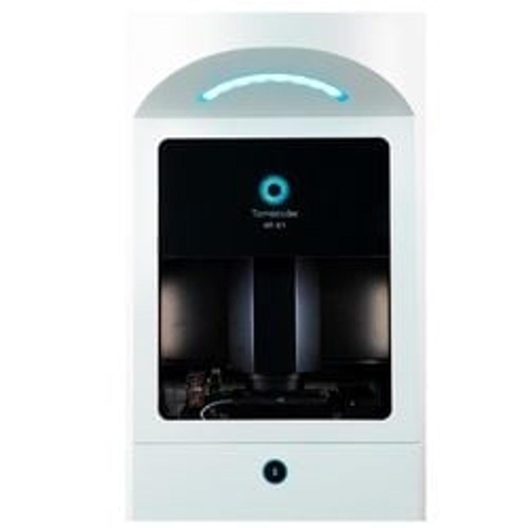
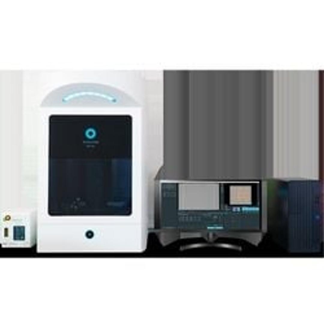
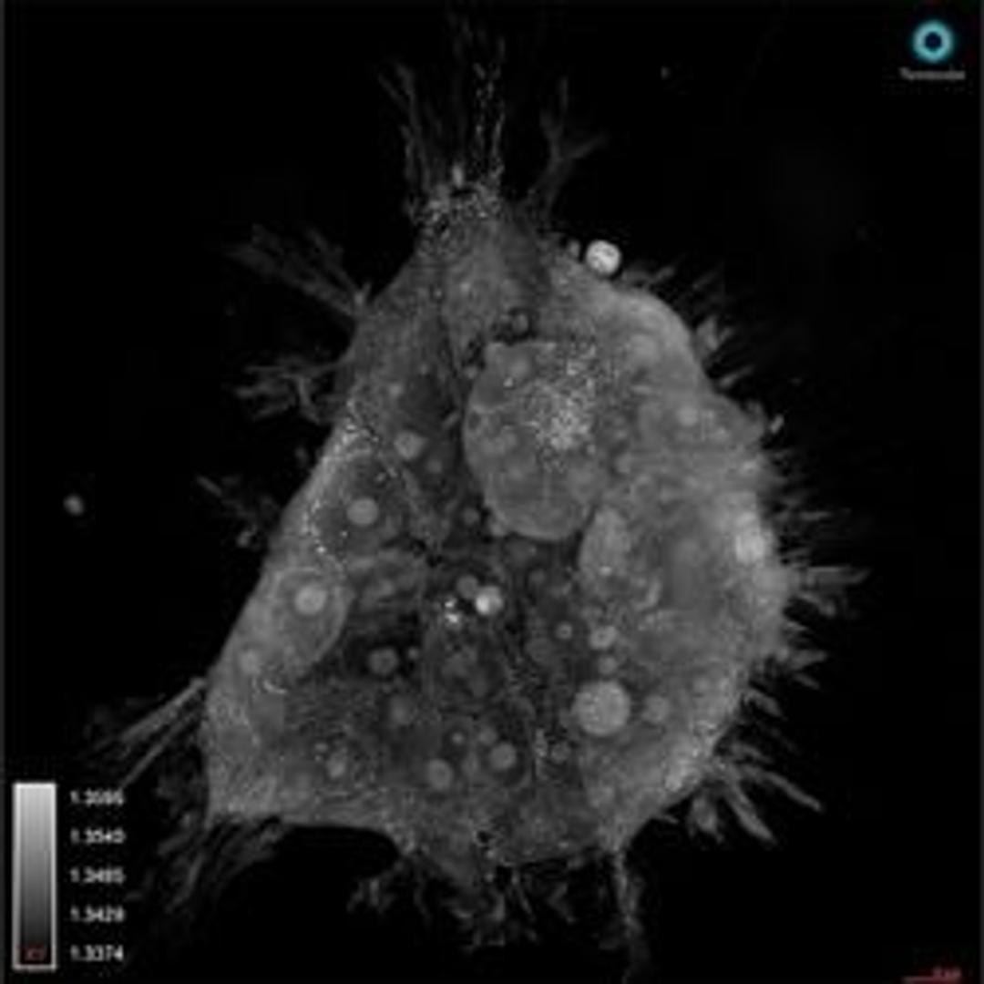
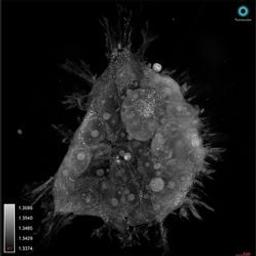
The Tomocube HT-X1 Holotomography Imaging System is a cutting-edge digital holotomography (HT) imaging system that enables label-free three-dimensional (3D) visualization of transparent specimens. The HT-X1 generates 3D refractive index (RI) tomograms representing the distribution of the RIs of specimens, which can then be translated into morphological, chemical, and mechanical properties.
The HT-X1 incorporates the high resolution, high contrast, and high sensitivity capabilities of the HT series from Tomocube and adapts them into a programmable multi-well plate imaging system with laser autofocus at a larger field of view. The HT-X1 is the first-ever holotomography technique to use a low-coherent light source with multiple beam patterns to obtain quantitative 3D RI information, thereby minimizing interference noise and eliminating the need for a calibration step for image acquisition. The motorized stage allows for tiling and multi-point analysis within each well, in addition to moving between wells. A built-in stage-top incubation system completes the live-cell imaging setup. A 3D fluorescence imaging module is incorporated to expand the information about the specimens by adding molecular information.
The HT-X1 has outstanding performance in the key applications of quantitative phase imaging, such as:
- Confluent & sensitive live cells: primary cells including stem cells, iPSC, neuronal cells, etc.
- Monitoring of multiple samples in a multi-well imaging plate
- Observing thicker samples: organoid, tissue, microfluidic device
- Nanomaterial delivery: better resolution without speckle noise


